
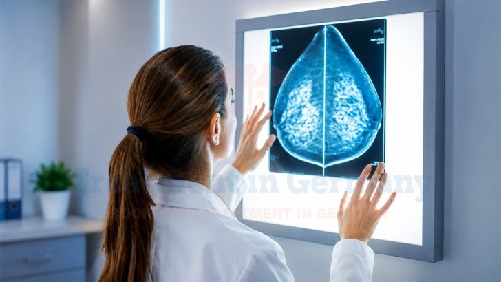
Breast cancer screening and diagnostic tests help doctors detect and evaluate breast abnormalities at different stages. This guide covers the main imaging methods, including mammography, ultrasound, and MRI, as well as biopsy and receptor testing used to confirm and characterize breast cancer. It also explains screening recommendations for different risk groups and the breast cancer diagnostic pathway in Germany.
Breast Cancer Diagnosis and Screening: Tests, Imaging and Biopsy
Breast cancer screening helps detect cancer at an early stage, sometimes before any noticeable symptoms appear. Mammography is the main screening test, while ultrasound and MRI may be used in certain situations depending on a person's age, breast density, and risk level. If screening or a physical examination shows something unusual, diagnostic tests can help doctors determine what the finding means. This may include additional imaging and, when necessary, a biopsy to confirm whether cancer is present.
Breast Cancer Screening and Early Detection

Breast cancer screening aims to find cancer early, often before symptoms develop, when more treatment options may be available [1].
A screening mammogram checks both breasts on a routine schedule in someone without symptoms. Diagnostic imaging, on the other hand, investigates a specific complaint [2].
An abnormal screening result does not confirm cancer on its own. It generally means:
- Additional imaging views may be needed to get a clearer picture.
- Another test, such as ultrasound, may be used to look closer.
- Tissue sampling (biopsy) may be recommended if the finding still looks suspicious.
- Most abnormal screening results turn out not to be cancer.
Screening recommendations depend on age and a person's individual risk. This means the recommended starting age and screening interval can differ between people.
Breast Cancer Screening in Germany: Standard and High-Risk Recommendations
Breast cancer screening in Germany varies according to age and individual risk. For women at average risk, the organized Mammography Screening Programme offers mammography every two years from ages 50 to 75. Women aged 30 and above can also receive an annual clinical breast examination. People with a higher or hereditary risk, including those with a BRCA1 or BRCA2 pathogenic variant or previous chest radiation at a young age, may need an individualized screening plan with additional imaging such as breast MRI and mammography. The exact starting age, interval, and combination of tests depend on the individual's risk and applicable guidelines. [10]

The table above summarizes the main breast cancer screening approaches in Germany for average-risk, higher-risk, hereditary-risk, and previous chest-radiation groups.
Screening for People at Higher Risk
Some people have a higher-than-average risk of breast cancer and may need to begin screening earlier or have additional imaging [3].
- A strong family history of breast or ovarian cancer.
- An inherited pathogenic variant such as BRCA1 or BRCA2.
- Previous chest radiation between roughly ages 10 and 30.
For people with a confirmed BRCA1 or BRCA2 pathogenic variant, high-risk screening may include regular breast MRI and mammography through an individualized, risk-adapted protocol, typically coordinated through a specialized high-risk breast cancer program. The exact schedule and starting age depend on the individual's risk and applicable guideline. Breast MRI is generally added to mammography rather than replacing it in these higher-risk groups. A gynecologist, breast specialist, or multidisciplinary hereditary breast and ovarian cancer center can assess the individual's risk and determine whether genetic counseling, earlier screening, or intensified imaging is appropriate.
Breast Cancer Screening Tests and Imaging

Several imaging methods examine breast tissue. Which test is used depends on factors such as age, breast density, personal risk, and whether doctors are screening for cancer or investigating a specific finding.
Mammography for Breast Cancer Screening
Mammography, an X-ray imaging test of the breast, remains the primary imaging method with the strongest population-level evidence for reducing breast cancer mortality [2]. A mammogram can detect:
- Masses or lumps in the breast tissue.
- Areas of distorted breast tissue.
- Calcifications: small calcium deposits, some of which are linked to early cancer.
Mammography's sensitivity varies with breast density. It performs less well in dense breast tissue, where overlapping fibroglandular tissue can obscure a small mass [4]. When a screening mammogram shows an abnormal finding, the next step is usually additional views or targeted ultrasound.
Breast Ultrasound for Screening and Evaluation
Breast ultrasound uses sound waves instead of radiation. It is especially useful for characterizing a finding, such as determining whether a mass is solid or fluid-filled. Supplemental ultrasound may detect additional cancers in some women with dense breasts, but it can also increase false-positive findings and lead to additional imaging or biopsies [4].
Dense breast tissue alone does not automatically mean supplemental MRI or ultrasound is required; recommendations vary according to overall risk and the guideline being followed. Ultrasound is also commonly used to evaluate breast findings in younger women and during pregnancy because it does not use ionizing radiation. Mammography can also be performed during pregnancy when clinically indicated. Ultrasound is not typically used alone for population-wide screening. In younger women or particular clinical situations, ultrasound may be the initial imaging test. It complements mammography rather than replacing it.
Breast MRI for Screening and Diagnosis
Contrast-enhanced breast MRI is among the most sensitive breast imaging methods available, particularly for women at elevated risk [4]. MRI is therefore mainly used for screening people with a high lifetime risk of breast cancer, particularly confirmed BRCA1 or BRCA2 carriers [3]. In selected cases, MRI can also help assess the extent of a newly diagnosed cancer or clarify a finding when mammography and ultrasound are inconclusive. MRI is not a routine, first-line screening test for average-risk women. Its high sensitivity comes with lower specificity, which can lead to extra imaging or biopsies for findings that prove benign.
Differences between Mammography, Ultrasound, and MRI
Mammography, breast ultrasound, and breast MRI have different roles in breast cancer screening and diagnosis. Mammography is the standard imaging test for routine screening and can detect breast masses and calcifications, although dense breast tissue can reduce its sensitivity. Breast ultrasound is mainly used to evaluate a specific breast finding, such as a lump or an abnormal mammogram, and does not use ionizing radiation. Breast MRI is more sensitive than mammography and is mainly used for screening people at high risk or for selected diagnostic situations, but it has lower specificity and may lead to additional tests or biopsies. Doctors may use more than one imaging method when additional information is needed. [4]

The table above compares the main uses, advantages, and limitations of mammography, breast ultrasound, and breast MRI.
Breast Cancer Diagnosis: From Imaging to Biopsy

A suspected breast abnormality may be noticed by the patient because of breast cancer symptoms, found during a clinical examination, or detected on a screening test. Doctors then use imaging and, when needed, biopsy to determine what the finding represents [5].
- Clinical assessment.
- Diagnostic imaging.
- Biopsy.
- Pathological analysis.
Clinical Breast Examination
A clinical breast examination involves a clinician visually inspecting and manually palpating the breasts and nearby lymph node areas for abnormalities. This can include a palpable lump, skin changes, or nipple discharge or retraction. Physical examination provides useful information, but it cannot, on its own, confirm or rule out cancer. Any suspicious finding on examination needs to be followed by imaging and, in most cases, tissue sampling before a diagnosis is established.
Diagnostic Breast Imaging
Diagnostic imaging differs from screening imaging in that it targets a specific area of concern rather than surveying the whole breast on a routine schedule. A diagnostic mammogram typically includes additional or magnified views, with targeted ultrasound frequently used alongside it to characterize the finding. Breast MRI may be considered in selected situations, for example to assess disease extent in a newly diagnosed cancer and help determine the breast cancer stage [4].
Breast Biopsy and Pathology
A biopsy, removal of a small tissue sample for laboratory analysis, is generally recommended when imaging shows a finding suspicious enough to warrant tissue confirmation [6].
Common biopsy approaches include:
- Core needle biopsy is commonly preferred for suspicious breast findings, with imaging guidance chosen according to how the lesion is best visualized.
- Mammographic (stereotactic) or MRI-guided biopsy is used less commonly, for findings only visible on those tests.
- Fine-needle aspiration is used selectively, for example for certain cystic or lymph-node findings.
- Surgical excisional biopsy is used in selected cases.
A pathologist examines the tissue to determine whether cancer is present and assesses features such as its histologic type and grade of breast cancer [7].
Pathology results are also checked against the imaging findings to ensure concordance. If the biopsy result does not adequately explain the imaging finding, the radiologist and pathologist may recommend repeat biopsy or, in some cases, surgical excision.
Breast Cancer Biomarker and Receptor Testing

Once breast cancer is confirmed on biopsy, the same tissue sample is typically tested further to characterize the tumor's biological features.
ER, PR and HER2 Testing
Estrogen receptor (ER), progesterone receptor (PR), and HER2 testing are routinely assessed in newly diagnosed invasive breast cancer [8] [12].
The results indicate:
- ER and PR status: whether hormones influence the tumor's growth.
- HER2 status: whether the tumor overexpresses the HER2 protein or carries HER2 gene amplification.
- How the cancer is classified, for example, hormone receptor-positive, HER2-positive breast cancer or triple-negative breast cancer [8].
ER, PR, and HER2 testing are standard parts of the diagnostic work-up. How each subtype is subsequently treated is beyond the scope of this diagnostic overview.
Additional Biomarker and Molecular Testing
For some patients with early-stage, hormone receptor-positive, HER2-negative breast cancer, additional multigene genomic assays may help estimate recurrence risk and inform adjuvant chemotherapy decisions [9].
These tests are not required for everyone and are used in selected clinical scenarios based on tumor biology, stage, lymph-node status, menopausal status, and other clinical factors. Genetic counseling and testing may be recommended when personal or family history suggests an inherited predisposition to breast cancer, with testing guided by the individual's risk and applicable guidelines [3].
Breast Cancer Screening and Diagnosis in Germany
Germany provides organized breast cancer screening alongside diagnostic services for people with symptoms or abnormal findings. Women aged 50 to 75 can take part in mammography screening every two years, while people with higher or hereditary risk may need an individualized screening plan with additional imaging [10].
How does breast cancer screening and diagnosis work in Germany?
Breast cancer screening generally uses mammography, while ultrasound or MRI may be added according to breast density, age, individual risk, or a specific finding. If screening or diagnostic imaging shows a suspicious abnormality, a biopsy may be performed to establish whether cancer is present and determine its pathological characteristics.
For international patients, assessment can begin with a review of previous imaging, pathology reports, and medical records before further tests are arranged.
Diagnostic costs depend on the imaging, biopsy, pathology, and other tests required. The cost of breast cancer treatment in Germany provides broader cost information, while the final diagnostic cost depends on the individual work-up.
TIG GmbH (Treatment in Germany) can help coordinate medical records, connect patients with appropriate breast specialists or certified breast centres, and arrange consultations where applicable. The treating medical team remains responsible for diagnostic decisions and clinical care.
References
1. American Cancer Society Early Detection and Diagnosis of Breast Cancer
2. US Preventive Services Task Force Screening for Breast Cancer: 2024 Recommendation Statement (JAMA)
3. NCCN Guidelines Genetic/Familial High-Risk Assessment: Breast, Ovarian, and Pancreatic
6. NCCN Clinical Practice Guidelines Breast Cancer Screening and Diagnosis (JNCCN)
7. National Cancer Institute Surgical Pathology Reports Fact Sheet
8. ASCO–College of American Pathologists Guideline Update HER2 Testing in Breast Cancer
10. Bundesamt für Strahlenschutz (BfS) The German Mammography Screening Programme
12. Estrogen and Progesterone Receptor Testing in Breast Cancer: ASCO/CAP Guideline Update
Why Patients Worldwide Prefer Our Medical Services in Germany – Key Benefits Explained
International patients planning breast cancer treatment in Germany may need support with hospital consultations, medical documentation, translations, and travel arrangements. TIG GmbH helps coordinate these practical steps and communication with healthcare providers according to each patient's needs and the services agreed.

The table above compares the support and coordination services offered through TIG GmbH with the arrangements patients may need to manage when booking directly with a clinic.

















.webp)
 (1).webp)

.webp)


.jpg)

 (1).webp)

.jpg)