

Breast cancer includes several types and subtypes that differ in where they develop, whether they invade surrounding tissue, and which biomarkers they carry. This guide explains DCIS, LCIS, invasive ductal and lobular carcinoma, hormone receptor-positive, HER2-positive, and triple-negative breast cancer, along with other less common forms and how these classifications can influence diagnosis and treatment.
Types of Breast Cancer: Invasive, Non-Invasive and Molecular Classification
Breast cancer includes several distinct types and subtypes. Doctors classify these cancers according to where they develop, whether they have invaded surrounding breast tissue, and which biological markers are present. These features are evaluated through breast cancer diagnosis and screening, pathology, and other clinical assessments, helping determine prognosis and treatment options. A pathology report may therefore describe a cancer using several terms, such as invasive ductal carcinoma or HER2-positive breast cancer.
Breast Cancer Classification by Type and Subtype

Breast cancer type usually refers to where the cancer began; subtype describes its biological or molecular characteristics. Together, a pathology report describes a tumor along several dimensions rather than a single classification:
- Histological origin: whether the cancer began in the milk ducts (ductal) or the lobules, the glands that produce milk (lobular).
- Invasive vs. non-invasive status: whether abnormal cells remain confined to their site of origin or have grown into surrounding breast tissue.
- Receptor characteristics: whether tumor cells express estrogen receptors (ER), progesterone receptors (PR), or HER2, which provides clinically important information about tumor biology. (Intrinsic molecular subtypes are defined separately, using gene-expression profiling.)
These traits provide complementary biological perspectives on the same malignancy, not competing classifications. Cancer can start in the lobules, invade surrounding tissue, and express hormone receptors. The pathology report lists histology, invasiveness, and biomarkers, such as invasive lobular carcinoma, ER-positive, PR-positive and HER2-negative.
Non-Invasive Breast Cancer Types

Non-invasive or in situ, conditions remain confined to the ducts or lobules where they originated. They have not broken through the surrounding basement membrane into nearby breast tissue, which distinguishes them from invasive disease.
Ductal Carcinoma in Situ (DCIS)
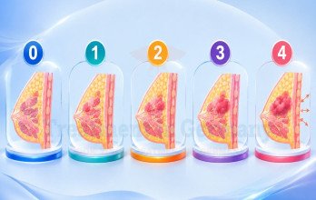
Ductal carcinoma in situ (DCIS) develops in the cells lining the milk ducts. The abnormal cells stay inside the duct wall, which is why DCIS is classified as Stage 0 within the broader framework of breast cancer stages and considered non-invasive disease [1].

DCIS accounts for approximately 20% of breast cancers, but this figure varies according to the demographic and screening setting. It cannot spread to lymph nodes or distant organs like invasive cancer because it remains in the ducts. DCIS does not metastasize; however, when DCIS is excised, an invasive component may be seen in the surgical specimen, which does carry a risk of spread.
Untreated DCIS lesions may progress to invasive breast cancer, although the chance varies by tumor and cannot be reliably predicted in advance. Grade, size, and necrosis may help estimate risk, but they cannot reliably predict which lesions will become invasive.
Lobular Carcinoma in Situ (LCIS)
Lobular carcinoma in situ (LCIS) consists of abnormal cells confined to the lobules, the glands that produce breast milk. Unlike DCIS, classic LCIS is generally considered a marker of increased future breast cancer risk rather than an invasive cancer itself [2].

Pathologists recognize three patterns, which differ in cell appearance and management:
- Classic LCIS is generally considered a marker of increased future breast cancer risk. Management is based on the individual's overall risk and may include regular breast screening, risk-reduction strategies, and, in selected patients, additional surveillance or preventive medication.
- Pleomorphic LCIS is often managed similarly to DCIS because its features are linked to a higher risk of coexisting invasive disease.
- Florid LCIS is also sometimes managed more like DCIS, for the same reason.
Invasive Breast Cancer Types

Invasive breast cancers have grown beyond their duct or lobule of origin into surrounding breast tissue, and from there can potentially reach the lymph nodes or bloodstream. Histological subtype is one of several factors alongside grade, size, and receptor status used to plan monitoring and treatment.
Invasive Ductal Carcinoma (IDC)
Invasive carcinoma of no special type (NST), commonly called invasive ductal carcinoma (IDC), is the most common histological type of invasive breast cancer. It originates in the milk ducts and grows through the duct wall into the surrounding breast tissue, accounting for roughly 70% to 80% of invasive breast cancers in commonly cited estimates [3].

Under the microscope, IDC may form cohesive nests, cords, or tubular structures that infiltrate the surrounding tissue. Clinically, it may present as a mass detected on mammography or physical exam. Grade, size, and lymph node involvement are assessed separately from histological type and are central to staging.
Invasive Lobular Carcinoma (ILC)
Invasive lobular carcinoma (ILC) begins in the breast lobules rather than the ducts. It's the second most common major histological type of invasive breast carcinoma, representing about 10% to 15% of invasive breast cancers [3].

A defining feature of classical ILC is loss of the cell-adhesion protein E-cadherin, which causes tumor cells to grow in single-file strands rather than forming a cohesive mass. This diffuse pattern can make ILC harder to detect on mammography and physical exam than IDC. ILC is also more likely than IDC to be hormone receptor-positive and HER2-negative [4], and its incidence has been steadily increasing over recent years compared to other breast cancer types.
Non-Invasive vs Invasive Types at a Glance
Breast cancer lesions differ in where they begin and whether they have invaded surrounding tissue. DCIS remains within the milk ducts and is classified as Stage 0 disease. Classic LCIS remains within the lobules and is mainly considered a marker of increased future breast cancer risk. IDC and ILC are invasive cancers that grow beyond their ducts or lobules. IDC is the most common invasive type, while ILC often has a single-file growth pattern.

The image above compares the main breast cancer types by origin, invasive status, and key features.
Breast Cancer Receptor and Molecular Subtypes

In addition to histological categorization, invasive breast cancer pathology commonly includes assessment of three receptors, ER, PR, and HER2 by immunohistochemistry and further testing if required, since these biomarkers are helpful in tumor classification and therapy guidance.
This receptor profile is useful for subtyping the tumor into clinically relevant subtypes. In the absence of gene-expression testing, doctors typically employ hormone receptor status, HER2 status, histological characteristics, and other indicators as clinical surrogates for intrinsic molecular subtypes, which are characterized by gene-expression profiling. Gene-expression-determined subgroups do not always correspond well to these clinical classifications. Receptor status is determined irrespective of whether the tumor is ductal or lobular.
Hormone Receptor-Positive Breast Cancer
HR-positive breast cancer expresses ER, PR, or both. Hormone-receptor status characterizes tumor biology and can suggest endocrine therapy benefit for ER-positive illness [6].

Hormone receptor status is particularly important when considering hormone and targeted therapy for breast cancer. Hormone receptor (HR)-positive tumors are classified into luminal A-like and luminal B-like subtypes. While these can be clinically approximated using ER, PR, HER2 and proliferation markers such as Ki-67, gene-expression profiling is required for definitive intrinsic molecular subtyping. Luminal A-like tumors proliferate less than B-like tumors. Hormone receptor-positive breast cancer, the most common receptor-based group, occurs in both IDC and ILC and has different molecular and histological characteristics [6].
HER2-Positive Breast Cancer
HER2-positive breast cancer is defined by HER2 protein overexpression and/or gene amplification identified through validated pathology testing. HER2 status is assessed primarily by immunohistochemistry (IHC), with in situ hybridization (ISH, including FISH) used in specific cases to clarify the result [9].

This subtype can occur with or without hormone receptor co-expression; hence, studies sometimes distinguish HER2-positive/HR-positive from HER2-positive/HR-negative illness. HER2 overexpression or amplification can change tumor biology and treatment selection, making it an important clinical biomarker in HER2-positive breast cancer. HER2-low is a treatment-related expression category rather than a distinct molecular subtype of breast cancer.
Triple-Negative Breast Cancer (TNBC)
Triple-negative breast cancer (TNBC) lacks expression of ER, PR, and HER2. It accounts for approximately 15% to 20% of all breast cancers, though estimates vary depending on the population and diagnostic criteria. While it closely overlaps with the basal-like molecular subtype identified by gene-expression profiling, the two are not synonymous.

TNBC is not one uniform disease; it is biologically heterogeneous, meaning that tumors with the same triple-negative receptor profile can still differ at the molecular level. These differences are an active area of research and may influence future treatment strategies [7].
Breast cancers can also be grouped by their biological characteristics. Luminal A-like tumors are generally hormone receptor-positive with lower proliferation, while luminal B-like tumors tend to show higher proliferation. HER2-positive cancers have HER2 overexpression or amplification and may also be hormone receptor-positive. Triple-negative breast cancer lacks ER, PR, and HER2 expression. These profiles help describe tumor biology and guide treatment decisions.

The image above summarizes the main breast cancer groups according to hormone receptor status, HER2, and biological characteristics.
Other and Rare Breast Cancer Types

Beyond invasive ductal and lobular carcinoma, a handful of rarer presentations round out the classification picture.
Inflammatory breast cancer (IBC) is a biologically distinct, aggressive form accounting for roughly 1–5% of cases. Rather than forming a distinct lump, it causes rapid swelling, redness, warmth, and a pitted, orange-peel skin texture (peau d'orange) as cancer cells block lymphatic vessels in the skin. IBC can occur with any hormone receptor or HER2 status, is classified as T4d, and is typically staged as III or IV depending on lymph node involvement and metastasis. Because these symptoms can mimic a common breast infection (mastitis), any sudden breast redness, swelling, warmth, or skin dimpling requires immediate medical evaluation, especially if symptoms do not improve rapidly with antibiotics [8].
A smaller group of special type invasive carcinomas make up the rest of diagnoses, with prognosis driven more by stage, grade, and receptor status than by histology alone. Mucinous and tubular carcinomas tend to have more favorable outcomes when caught early, while carcinoma with medullary features and metaplastic carcinoma are more often high-grade and triple-negative.
Other uncommon breast cancer presentations include Paget disease of the breast, which affects the nipple and areola and is usually linked to an underlying DCIS or invasive cancer. Persistent nipple changes such as itching, redness, scaling, or crusting that does not heal warrant prompt medical evaluation to rule out Paget disease. Phyllodes tumors are fibroepithelial growths ranging from benign to malignant, and both are managed separately from the ductal, lobular, and molecular subtypes above [10].
Because these classifications shape treatment decisions, an unclear or unusual diagnosis may benefit from review at a specialty breast center. For a broader overview of breast cancer treatment in Germany, including treatment approaches and specialist care, patients can explore the available options through TIG GmbH (Treatment in Germany).
References
1. American Cancer Society: Ductal Carcinoma In Situ (DCIS)
2. American Cancer Society: Your Breast Pathology Report: Lobular Carcinoma In Situ (LCIS)
3. NCI SEER Cancer Stat Facts: Invasive Lobular Carcinoma
5. Giaquinto et al., Cancer (American Cancer Society) Lobular Breast Cancer Statistics, 2025 (PMC)
6. NCI Breast Cancer Biomarker Tests Hormone Receptors, HER2, and Others
7. Cancer Discovery (AACR) Insights into Molecular Classifications of Triple-Negative Breast Cancer
8. NCBI StatPearls Inflammatory Breast Cancer
9. ASCO/CAP HER2 Testing in Breast Cancer: Guideline Update
Why Patients Worldwide Prefer Our Medical Services in Germany – Key Benefits Explained

















.webp)
 (1).webp)

.webp)


.jpg)

 (1).webp)


.jpg)