
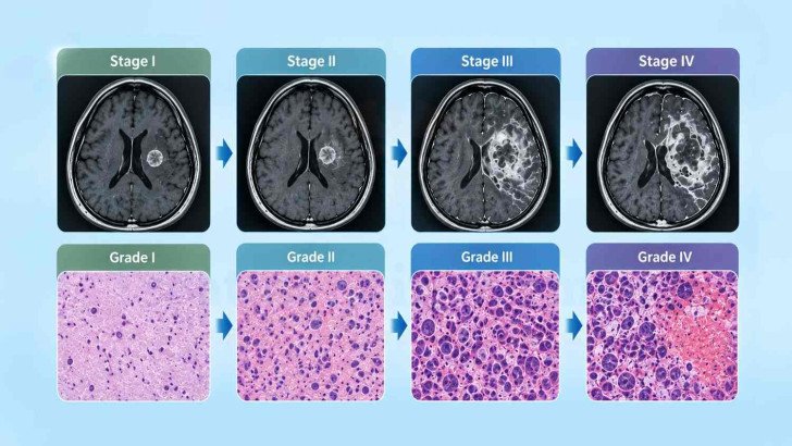
Most primary brain tumors are classified by tumor type, CNS WHO grade, molecular findings and disease extent rather than conventional TNM stage. Grade reflects biological behavior, while location and extent describe where the tumor is and how much of the central nervous system is involved. Modern WHO classification combines histology with molecular findings, and the final diagnosis may be refined after surgery, molecular testing or specialist neuropathology review.
Brain Cancer Stages and Grades: WHO Classification, Grade and Tumor Extent
Most primary brain tumors are not assigned a conventional cancer stage. Instead, the diagnosis describes the tumor type, CNS WHO grade, molecular findings and disease extent. Each provides different information and should be interpreted together. These factors also help guide decisions about brain cancer treatment in Germany, including the role of surgery, radiation therapy and drug treatment.
Brain Tumor Disease Extent and Staging
Cancer staging systems generally describe the extent of a tumor at its original site, involvement of regional lymph nodes and spread to distant organs. This forms the basis of the TNM system used for cancers such as breast, lung and colorectal cancer. Primary brain tumors behave differently because they rarely spread outside the central nervous system and do not typically spread through regional lymph nodes. For this reason, most primary adult brain tumors are not assigned a TNM stage.
Tumor size alone does not adequately describe the clinical significance or prognosis of a primary brain tumour. For example, a three-centimetre tumor in the frontal region and a one-centimetre tumor affecting the brainstem may have very different clinical implications because of their location. Clinicians therefore also describe:
- Location, including the anatomical structures involved, displaced or invaded by the tumour.
- Whether it crosses the midline, involves both hemispheres or reaches the deep central structures.
- Whether disease is multifocal, appearing in more than one place at once rather than as a single mass.
- Whether tumor cells have reached the cerebrospinal fluid, which matters particularly for tumour types such as medulloblastoma and other CNS embryonal tumours,
- Residual tumor remaining after surgery, which is reported in defined categories rather than as a stage [1].
There is one common exception. Tumors that do spread through the spinal fluid, medulloblastoma being the usual example in children, are assessed for that spread and the results are incorporated with postoperative residual disease, tumour biology and patient age to determine risk group. Brain metastases are secondary tumours rather than primary brain cancers and are classified according to the staging system for their primary cancer. For example, a lung cancer that has reached the brain is staged as advanced lung cancer.
These findings do not usually result in a conventional numerical stage. Instead, tumor location and extent are assessed separately because both can significantly influence symptoms, treatment options and clinical management.
Understanding Brain Tumor Grade and Aggressiveness
Tumor grade describes the expected biological behavior of a tumor, including how rapidly it is likely to grow and, for some tumor types, its tendency to infiltrate surrounding tissue. Grading is based on microscopic features assessed by a neuropathologist and, under current classifications, may also incorporate specific molecular findings.
Microscopic features considered during tumor grading include:
- Cell density: or how tightly the tumor cells are packed together in the tissue.
- Atypia: meaning how abnormal and irregular the individual cells look.
- Mitotic activity: the number of cells caught in the act of dividing when the sample was taken.
- Microvascular proliferation: where the tumor has driven new and disordered blood vessels to form.
- Necrosis: areas of dead tissue where growth has outstripped its own blood supply.
Broadly, the relative importance of these features depends on the specific tumour type, and CNS WHO grading is not determined simply by counting how many adverse microscopic features are present. For many tumour entities, molecular findings are integrated with histopathology to establish the final diagnosis and grade.

What each CNS WHO grade broadly indicates. Grade is assigned within a tumor type, so the same number carries different weight across different diseases.
Grades 1 and 2 are commonly described as low-grade, while grades 3 and 4 are described as high-grade. However, this distinction is only a general guide because grade is assigned within a specific tumor entity rather than across all brain tumors. Grade also does not describe tumor location, which can have a major influence on symptoms and treatment options.
Modern WHO Classification and Molecular Features
For much of the twentieth century, brain tumors were classified primarily according to their microscopic appearance. Although this approach provided important diagnostic information, tumors with similar histological appearances could have substantially different biological behavior and clinical outcomes.
The fifth edition of the WHO Classification of Tumours of the Central Nervous System, published in 2021 and commonly referred to as WHO CNS5, incorporated molecular findings directly into the classification of many CNS tumors. Diagnosis may therefore integrate two main sources of information:
- Histology: cell appearance, tissue architecture, mitotic activity and features such as necrosis or microvascular proliferation
- Molecular findings: depending on the tumor type, these may include IDH mutation status, 1p/19q codeletion, CDKN2A/B deletion, TERT promoter mutation and, in selected centres, DNA methylation profiling.
Combining these findings produces an integrated diagnosis, sometimes referred to as a histomolecular diagnosis. For example, a report may state: astrocytoma, IDH-mutant, CNS WHO grade 3. Each component provides specific diagnostic information.
Molecular findings can also directly influence tumor grade. In IDH-mutant astrocytoma, homozygous deletion of CDKN2A/B is sufficient for classification as CNS WHO grade 4 even when necrosis or microvascular proliferation is absent [2]. This illustrates how current grading may be determined by molecular characteristics as well as microscopic appearance.
The effect of this change can be seen in clinical practice. When a Beijing centre re-examined 452 gliomas treated over an eleven-year period and reclassified them according to WHO CNS5, the tumors were categorized as adult-type diffuse gliomas (373), pediatric-type diffuse gliomas (23), circumscribed astrocytic gliomas (20), and glioneuronal and neuronal tumors (36), with the distribution differing from classifications based on earlier criteria. Older pathology reports may therefore contain terminology that has since been revised or retired but was appropriate under the classification criteria used at the time [3].
Two abbreviations turn up in reports and are easily confused:
- NOS: not otherwise specified, means the molecular information needed for a full diagnosis was never available, usually because the testing was not done or the sample was insufficient.
- NEC: not elsewhere classified, means the testing was completed and the results simply do not match any entity the classification currently recognizes.
Brain Tumor Grade, Extent and Clinical Classification
Theimportance of interpreting tumor type together with grade is illustrated by two tumors that both carry CNS WHO grade 4. In a University of Virginia cohort, 972 patients with glioma were screened, and 736 had sufficient data for classification under WHO CNS5. Patients with grade 4 IDH-mutant astrocytoma presented at a mean age of 39.8 years, compared with 63.1 years for patients with IDH-wildtype glioblastoma [4].

Mean age at presentation for two tumors that both carry CNS WHO grade 4, from a cohort of 736 patients classified under CNS5 [4].
Clinical interpretation also considers tumor location, residual tumor after surgery, relevant molecular findings and changes over time on serial imaging. These factors are assessed alongside tumor type and grade because no single feature provides a complete picture of the disease.
Tumor grade represents only one component of the diagnosis. Patients may therefore benefit from knowing the complete integrated diagnosis, including tumor type, grade and relevant molecular findings.
When Brain Tumor Classification Changes
A brain tumor diagnosis may be refined after the initial pathology assessment as additional diagnostic information becomes available. The final terminology may change for several reasons:
- More tissue becomes available: A resection specimen shows more than a needle biopsy, and features absent from the small sample may be present in the larger one.
- Molecular results arrive: These commonly lag the microscopic report by days or weeks and can alter both the tumor name and the grade.
- A specialist neuropathologist reviews the case: Difficult tumors are routinely sent out for expert opinion.
Criteria are updated. A tumor diagnosed before 2021 may be reclassified under current criteria without any biological change in the tumor itself.
Specialist neuropathology review can be particularly valuable in diagnostically challenging cases. In a study of 141 selected pediatric CNS tumors from a hospital in Karachi that were reviewed at a specialist centre in Toronto, the reviewing pathologists disagreed with the original diagnosis in 30 cases and provided a more precise subclassification in 53 cases. These findings apply to a selected group of difficult cases rather than to all brain tumor diagnoses [5].
It is important to distinguish diagnostic reclassification from disease progression. Reclassification means that the understanding or terminology of the tumor has been refined and does not, by itself, indicate a biological change in the tumor. Progression, by contrast, refers to a change in the disease itself.
The final integrated diagnosis may take longer than the initial pathology report, particularly when molecular testing or external specialist review is required. The additional time allows histological and molecular findings to be integrated into a more precise diagnosis.
References



































.webp)
 (1).webp)

.webp)


.jpg)

 (1).webp)


.jpg)