

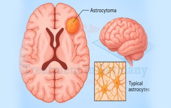
Astrocytoma and diffuse glioma treatment in Germany depends on tumor type, grade, molecular findings, extent of surgery, symptoms and overall health. Care may involve surgery, biopsy, active surveillance, radiotherapy, chemotherapy, molecularly guided treatment, follow-up and treatment after progression. Specialist review in Germany can help clarify difficult diagnoses, residual disease, treatment choices and the role of newer options such as vorasidenib.
Astrocytoma and Glioma Treatment in Germany: From Surgery to Long-Term Care
Two people diagnosed with diffuse glioma may need very different treatment plans depending on the tumor’s integrated diagnosis, WHO grade, molecular characteristics, clinical features and treatment history. One may undergo surgery followed by MRI surveillance, while another may need radiotherapy and chemotherapy depending on the integrated tumor diagnosis and individual risk factors. Glioma treatment in Germany is tailored to the specific tumor type, CNS WHO grade, molecular findings, extent of tumor removal and the patient’s clinical condition rather than on the term “glioma” alone.
Astrocytoma and Glioma Treatment Planning
Before treatment decisions are made, brain tumor diagnosis involves combining histological and molecular findings to define the tumor more precisely. This is important because astrocytoma, oligodendroglioma and glioblastoma are biologically distinct diseases and may require different treatment approaches.
The multidisciplinary team also considers:
- tumor location and extent, particularly its relationship to areas involved in speech, movement or other critical functions.
- how much tumor remains after surgery.
- neurological symptoms and day-to-day function.
- age, general health and ability to tolerate treatment.
- previous treatment, where relevant.
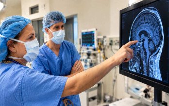
- whether follow-up MRI demonstrates stable disease, treatment-related changes, or findings consistent with tumor progression.
These factors are considered together rather than individually when deciding between surveillance, additional treatment or further intervention.
Surgery and Tumor Removal for Astrocytoma and Glioma
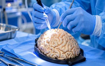
Brain tumor surgery is often the first major step because it provides tissue for histological and molecular diagnosis and, where safely possible, reduces tumor burden. The extent of surgery is determined by the tumor’s location and the need to preserve neurological function, where the goal is maximal safe resection rather than removal at the expense of an neurological deficit.
Maximal Safe Tumor Removal
When removal is possible without an unacceptable risk of neurological damage, the surgical goal is maximal safe resection. However, diffuse gliomas infiltrate surrounding brain tissue, so an MRI showing no visible residual tumor does not mean that all microscopic tumor cells have been removed.
Postoperative management therefore depends on more than the extent of resection. The integrated diagnosis, neurological status and postoperative imaging help determine whether surveillance or additional treatment is appropriate [1].
Biopsy When Tumor Removal Is Not Suitable
When tumor removal would carry an unacceptable neurological risk, stereotactic biopsy may be used to obtain tissue for diagnosis. This may be appropriate for deep, highly infiltrative or functionally critical tumors.
Biopsy usually provides enough tissue for histological and molecular assessment, although diagnostic yield is not 100%. In a series of 1,214 stereotactic biopsies for suspected glioma, an integrated diagnosis was obtained in 96.3% of procedures [4]. Unlike resection, biopsy does not substantially reduce tumor volume.
Active Surveillance for Selected Astrocytoma and Glioma
For selected patients, active surveillance may be appropriate after surgery when immediate radiotherapy or chemotherapy can reasonably be deferred. This is a planned management strategy involving scheduled MRI and clinical assessment rather than a delay in care.
Surveillance may be considered when:
- the integrated diagnosis indicates more favorable tumor biology.
- no visible tumor or only stable residual disease remains after surgery.
- neurological symptoms are absent or well controlled.
Because glioma growth can vary between patients, changes are assessed over serial scans rather than from a single measurement. Treatment may be reconsidered if there is convincing tumor growth, worsening neurological symptoms, new contrast enhancement or clinically important new pathological or molecular findings [5].
Radiation and Drug Treatment for Astrocytoma and Glioma
Radiotherapy and systemic treatment are selected according to the integrated diagnosis, CNS WHO grade, molecular findings and clinical condition. They are not required after every glioma diagnosis.
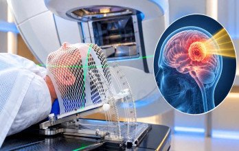
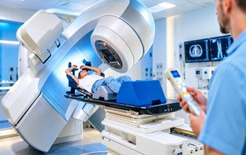
When treatment after surgery is needed, the regimen depends on the tumor type. IDH-mutant, 1p/19q-codeleted oligodendroglioma is commonly treated with radiotherapy and PCV chemotherapy. CNS WHO grade 3 IDH-mutant astrocytoma is generally treated with radiotherapy followed by adjuvant temozolomide. For CNS WHO grade 2 IDH-mutant astrocytoma that requires active treatment, radiotherapy with adjuvant chemotherapy is recommended. For selected patients with CNS WHO grade 2 IDH-mutant astrocytoma or oligodendroglioma, radiotherapy and chemotherapy may be deferred. Surveillance or, when appropriate, vorasidenib may then be considered [10].
For glioblastoma, treatment commonly involves surgery when feasible, followed by radiotherapy and temozolomide chemotherapy, depending on the patient's clinical circumstances. Further details are available in our article on glioblastoma treatment in Germany.
Molecular Factors Affecting Astrocytoma and Glioma Treatment
Molecular findings help define the tumor type and grade and can also influence treatment decisions. They are interpreted as part of the integrated diagnosis rather than in isolation [1] [2] [3].
Molecular testing can change the diagnosis. Under the current WHO classification, oligodendroglioma requires both an IDH mutation and 1p/19q codeletion. Some tumors previously called oligodendroglioma therefore no longer meet the current definition [6]. Older terms such as “anaplastic astrocytoma” and “oligoastrocytoma” may also not correspond directly to current tumor categories [2] [6].
Treatment Differences Across Lower- and Higher-Grade Gliomas
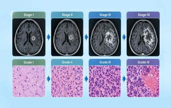
Brain tumor grades and WHO classification help describe tumor biology, but treatment decisions also depend on the integrated histomolecular diagnosis, molecular alterations, tumor location and extent, patient factors, and prior treatment. Molecularly defined glioma types can behave differently even within the same grade [3]
Selected patients with CNS WHO grade 2 IDH-mutant glioma may initially undergo surveillance or, when eligible, receive vorasidenib, whereas grade 3 and grade 4 tumors generally require more intensive treatment. For this reason, terms such as “low-grade” and “high-grade” rather than as substitutes for the specific integrated histomolecular diagnosis [7] [10].
Follow-Up After Astrocytoma and Glioma Treatment
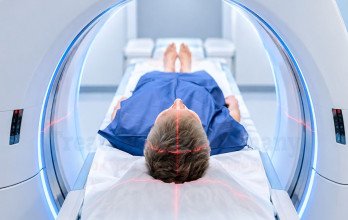
Long-term follow-up is required after both active treatment and surveillance because diffuse gliomas can recur or progress. It usually includes MRI, neurological examination, symptom review and assessment of daily functioning, with the frequency determined by the diagnosis, previous treatment and clinical course.
Rehabilitation is based on individual needs and may include physiotherapy, occupational therapy, speech and language therapy or neuropsychological support. Patients referred for rehabilitation after brain tumor surgery may require multidisciplinary support [9].
Evaluating Astrocytoma and Glioma Progression
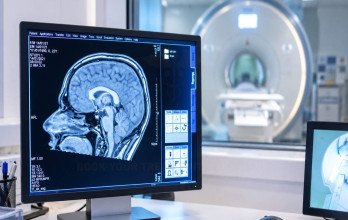
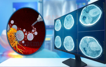
A new MRI abnormality does not always indicate tumor progression. Surgery and radiotherapy can produce treatment-related changes that may resemble tumor growth, so findings are interpreted alongside previous scans, symptoms and treatment history. Repeat or additional imaging may be required when progression remains uncertain and additional imaging such as perfusion MRI or amino-acid PET may be considered; where the appropriate approach depends on the clinical and treatment context [1].
Treatment After Astrocytoma and Glioma Progression
When progression is confirmed, treatment is reconsidered according to tumor location, neurological condition and previous treatment. Options may include repeat surgery, re-irradiation, further systemic treatment, molecular reassessment when the result could affect treatment, a clinical trial, or symptom-directed supportive care.
There is no single treatment sequence for recurrent diffuse glioma. Management depends on the pattern of progression, previous treatment, available options and the patient’s goals [1] [2].
Emerging Treatments for Astrocytoma and Glioma
Vorasidenib is an oral IDH1/IDH2 inhibitor for selected patients with CNS WHO grade 2 IDH-mutant astrocytoma or oligodendroglioma after surgery when radiotherapy and chemotherapy can be deferred. In the phase 3 INDIGO trial of 331 patients, median progression-free survival was 27.7 months with vorasidenib compared with 11.1 months with placebo. Current ASCO-SNO guidance includes vorasidenib as an option for eligible patients in this setting [8] [10].
Other emerging approaches include treatments selected for specific molecular changes, newer methods of delivering drugs to the brain, vaccine and cell-based approaches, immune-based treatments, tumor treating fields, and combination strategies. Their role varies because the strength of evidence, availability and suitability for individual patients differ between approaches. Some may be considered only in selected clinical settings.
Specialist Astrocytoma and Glioma Treatment in Germany
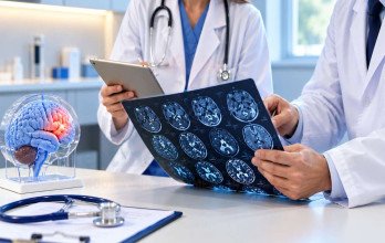
Diffuse glioma treatment in Germany is commonly coordinated through university hospitals and specialist neuro-oncology centres. Neurosurgery, neuropathology, neuroradiology, radiation oncology and medical oncology may review the diagnosis and treatment plan together. Integrated molecular pathology is an important part of this process because the final tumor classification can directly affect treatment.
Specialist review may be particularly useful when the diagnosis or molecular classification is uncertain, the tumor involves a surgically difficult or functionally important area, or a second opinion is needed before major treatment.
Review typically considers imaging, pathology, molecular findings and previous treatment history to clarify the diagnosis and available treatment options. Services and expertise may differ between centres.
TIG GmbH (Treatment in Germany) can coordinate the process for patients seeking specialist glioma care in Germany, including medical-record transfer, specialist review, hospital communication, appointment coordination and the practical arrangements needed for the next steps in care.
References
Why Patients Worldwide Prefer Our Medical Services in Germany – Key Benefits Explained
International patients planning breast cancer treatment in Germany may need support with hospital consultations, medical documentation, translations, and travel arrangements. TIG GmbH helps coordinate these practical steps and communication with healthcare providers according to each patient's needs and the services agreed.

The table above compares the support and coordination services offered through TIG GmbH with the arrangements patients may need to manage when booking directly with a clinic.











.webp)
 (1).webp)
.webp)
.jpg)
 (1).webp)
.jpg)