
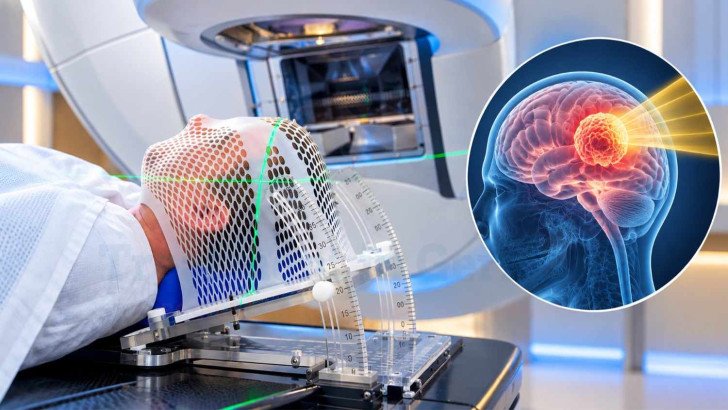
Stereotactic radiosurgery (SRS) uses highly focused radiation to treat selected brain tumors and metastases without surgical removal. Suitability depends on tumor type, size, location, number of lesions, previous treatment and overall health. Treatment may be given in one or several sessions using Gamma Knife, CyberKnife or LINAC-based systems. Recovery is usually quick, but swelling, fatigue and delayed radiation effects can occur, so follow-up MRI is important.
Stereotactic Radiosurgery for Brain Tumors in Germany: Suitability, Process and Outcomes
Stereotactic radiosurgery (SRS) is not a surgical operation. It uses highly focused radiation to treat a defined target inside the brain without an incision or surgical removal of tissue. Most patients are awake during treatment, although local anaesthetic may be used when a rigid head frame is required. Stereotactic radiosurgery in Germany is planned according to the tumor’s size, location, type and the patient’s overall condition.
Stereotactic Radiosurgery Uses in Brain Tumor Treatment
Stereotactic radiosurgery uses multiple highly focused radiation beams or arcs to deliver a high dose to a small, precisely defined target while limiting radiation exposure to surrounding brain tissue. Because SRS is a local treatment, it is best suited to discrete, radiographically well-defined targets rather than diffuse microscopic disease. It does not treat diffuse microscopic tumor spread beyond the target, which limits its role in infiltrative brain tumors.
SRS may be used for:
- Selected well-defined intracranial tumors, including some meningiomas and schwannomas particularly when the target size, location and clinical circumstances are appropriate.
- Brain metastases in appropriately selected patients, taking into account lesion number, total tumor volume, location and performance status [1].
- Selected residual or recurrent tumors or residual disease forms a sufficiently defined target and the prior treatment history.
- Postoperative cavities after resection of brain metastases.
Unlike surgery, SRS does not remove tissue or provide a specimen for pathological diagnosis. Conventional radiotherapy is usually delivered over more treatment sessions, while fractionated stereotactic radiotherapy uses the same highly focused approach over several sessions. The treatment goal depends on the diagnosis and may include durable local control, prevention of local recurrence or symptom control.
Patient Eligibility and Selection for Brain SRS

Eligibility for SRS is based on several clinical and imaging factors rather than a single tumor-size threshold.
These include:
- Tumor type and whether the lesion is well defined.
- Tumor size and total treatment volume.
- Number and distribution of intracranial lesions.
- Proximity to structures such as the optic nerves, optic chiasm, brainstem and cochlea.
- Neurological symptoms and the presence of mass effect.
- General health and performance status.
- Previous surgery or brain radiotherapy.
- The extent and control of disease outside the brain in patients with metastases.
Tumor volume and location are particularly important. A lesion may be suitable for SRS in one location but require a different approach if it lies close to a radiation-sensitive structure. In a retrospective study of 332 brain metastases, treatment dose and gross tumor volume were independently associated with the risk of radiation necrosis [3]. Lesions near critical structures, previously irradiated areas and patients with a high intracranial tumor burden generally require multidisciplinary assessment involving radiation oncology, neurosurgery and neuroradiology.
SRS Planning, Treatment Delivery and Fractionation
The accuracy of SRS depends on detailed imaging, immobilisation, target definition, treatment planning and quality assurance before radiation is delivered.
Planning usually includes:
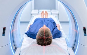
- High-resolution MRI and, for many treatment systems, a planning CT; the images are registered together when required.
- Immobilisation with a rigid stereotactic frame or a closely fitted thermoplastic mask, depending on the treatment platform.
- Delineation of the target and nearby critical structures.
- Dose planning to achieve high target coverage with a rapid dose fall-off outside the treatment area.
- Quality-assurance checks before treatment.
On the treatment day, the patient's position is verified according to the system being used. Treatment time varies with the platform, number and size of targets and complexity of the plan. Radiation itself is painless, and most adult patients remain awake during treatment.
Single-Session and Fractionated SRS
Single-fraction SRS delivers the prescribed dose in one treatment session and is commonly used for small, well-defined lesions when normal-tissue dose constraints can be met. Fractionated stereotactic radiotherapy delivers treatment over several sessions, commonly three to five. Fractionation can reduce the dose delivered to nearby normal tissue in each session and is often considered for larger lesions or targets close to critical structures. The schedule depends on tumor size, volume, location, previous radiotherapy and the dose limits of surrounding structures.
SRS Technologies and Delivery Platforms
SRS can be delivered using several treatment platforms. The most appropriate system depends on the target, treatment plan and expertise available at the treating centre.

The main delivery platforms. Each can deliver focused radiation accurately; none is universally superior.
No single SRS platform is best for every patient. Treatment quality depends on imaging, target delineation, dose planning, quality assurance and specialist experience. German centres may use dedicated radiosurgery systems or stereotactic-capable linear accelerators, and some offer more than one platform. Patients can ask which system is proposed and why it is appropriate for their lesion [2] [4].
SRS for Primary, Metastatic and Recurrent Brain Lesions
The role of SRS depends on the underlying diagnosis and previous treatment.
For selected well-defined intracranial tumors, SRS may provide durable local control. For brain metastases, SRS allows individual lesions to be treated while limiting radiation exposure to uninvolved brain tissue. Current guidelines recommend SRS for many patients with a limited number of brain metastases and good performance status, while whole-brain radiotherapy remains appropriate in selected situations [1]. Postoperative stereotactic radiotherapy can be used after resection of brain metastases to reduce the risk of recurrence in the surgical cavity. In a retrospective German study of 105 patients treated with fractionated stereotactic radiotherapy, estimated local control at one year was 98.6%. Local progression occurred in 2.0% during follow-up, while radiation-induced contrast enhancement was reported in 20.4%, at a median of 14.3 months [4].

Outcomes after fractionated stereotactic treatment of resection cavities in 105 patients [4]. High local control, with imaging changes appearing later in a fifth of patients.
Previous brain radiotherapy requires additional assessment because normal brain tissue may already have received a substantial radiation dose. Specialists review the previous dose distribution, interval since treatment, current target size and location, and the degree of overlap with previously irradiated tissue. Reirradiation may be possible in selected patients, but the original radiation plans are usually needed to assess cumulative dose and risk.
Side Effects, Recovery and Follow-Up After SRS
SRS is generally well tolerated, and many patients are discharged on the day of treatment. Early effects can include:
- Fatigue.
- Headache or discomfort related to immobilisation.
- Nausea.
- Temporary worsening of neurological symptoms because of swelling around the treated lesion.
- Seizures may occur, particularly in patients whose underlying brain lesion or clinical history places them at risk.
Peritumoral swelling may require corticosteroid treatment when symptomatic. A less common but clinically important delayed effect is radiation necrosis, which can develop months or sometimes years after treatment and may resemble tumor progression on MRI. Risk varies with factors including radiation dose, treated volume and previous radiotherapy [3] [2]. Many patients return to usual daily activities within a short period, although recovery depends on the underlying brain condition, symptoms and treatment received. Follow-up includes clinical assessment and repeat MRI.
Understanding Post-SRS Imaging
MRI appearances after SRS do not always reflect a simple reduction in tumor size. A treated lesion may temporarily enlarge or show increased contrast enhancement because of treatment-related changes, including radiation injury. These appearances can be difficult to distinguish from tumor progression. Interpretation therefore considers the timing of the scan, the patient's neurological condition and changes across serial MRI examinations. When conventional MRI remains uncertain, additional imaging techniques or multidisciplinary review may be required.
Specialist Stereotactic Radiosurgery in Germany

SRS in Germany is available through university hospitals and specialist centres where radiation oncology may work with neurosurgery, neuroradiology and neuro-oncology. Multidisciplinary assessment can be particularly important when a lesion is close to critical structures, when previous brain radiation has been given, or when more than one treatment option is being considered. The SRS platform available varies between centres. Depending on the institution, treatment may be delivered using Gamma Knife, CyberKnife or a LINAC-based system. The specific technology, relevant expertise and current treatment availability should therefore be confirmed directly with the individual centre.
How Can I Get a Specialist Opinion on SRS in Germany?
A specialist review can assess the tumor type, size, location, lesion burden and previous treatment to determine whether SRS may be appropriate. Recent MRI images and relevant medical records can often be reviewed before travel. If previous brain radiation has been given, the earlier radiation plans may also be required.
TIG GmbH (Treatment in Germany) can help international patients facilitate medical-record and imaging review, connect suitable cases with relevant radiation oncology specialists, and coordinate specialist consultations where applicable. The German medical team remains responsible for treatment assessment, recommendations and clinical care.
References
Why Patients Worldwide Prefer Our Medical Services in Germany – Key Benefits Explained
International patients planning breast cancer treatment in Germany may need support with hospital consultations, medical documentation, translations, and travel arrangements. TIG GmbH helps coordinate these practical steps and communication with healthcare providers according to each patient's needs and the services agreed.

The table above compares the support and coordination services offered through TIG GmbH with the arrangements patients may need to manage when booking directly with a clinic.


































.webp)
 (1).webp)
.webp)

.jpg)

 (1).webp)
.jpg)