

Brain tumors include gliomas, meningiomas, astrocytomas, oligodendrogliomas, glioblastomas and other non-glial tumors. Their diagnosis depends on microscopic appearance, molecular findings and tumor behavior. Primary tumors are different from brain metastases, and modern classification also considers grade and molecular features. Older pathology reports may use different names because brain tumor classification has changed over time.
Brain Cancer Types: Major Tumor Types and Classification
Two people can both be told they have a brain tumor and share almost nothing medically. One has an operation and needs nothing further. The other is told surgery is not an option. What separates them is the tumor type: what it looks like under the microscope, and the molecular changes it carries. These differences also influence the choice of brain cancer treatment in Germany, including whether surgery, radiation therapy, drug treatment or monitoring is appropriate.
Main Types of Brain Tumors
Where the tumor started is one of the first distinctions doctors make whether a tumor originated within the central nervous system or represents cancer that has spread to the CNS from another part of the body. Primary brain tumors begin in the brain or nearby structures. Secondary brain tumors, or brain metastases, spread there from cancer elsewhere in the body. They keep the identity of the original cancer: breast cancer that spreads to the brain is still classified and treated as metastatic breast cancer although treatment must also account for the characteristics and location of the brain metastasis.
Among primary tumors, grouping now follows a combination of microscopic appearance and molecular features with some tumor entities defined by specific molecular alterations.
Families then diverge along several lines at once:
- Their microscopic and molecular characteristics.
- Whether the tumor infiltrates brain tissue or presses against it from the outside.
- Age, since a few types are almost unheard of in adults and others almost never appear in children.
- The anatomical site or CNS compartment in which it characteristically arises.
- How often it turns out to be malignant, which ranges from rarely to nearly always.

The major families of primary brain and central nervous system tumors. Several smaller groups exist beyond these.
How common each family is varies enormously. Registry data from the United States covering 2017 to 2021 put meningioma at 41.7% of all primary brain and central nervous system tumors, with gliomas of every kind together making up 22.9% [1]. Outside the table sit smaller categories, among them glioneuronal and neuronal tumors, choroid plexus tumors and the mesenchymal group, some containing no more than a handful of recognized entities and several described for the first time only within the last decade or so.
Major Glioma Types in Brain Cancer

Glioma is a broad family rather than a single diagnosis. Modern classification separates several biologically distinct groups, including adult-type diffuse gliomas, pediatric-type diffuse gliomas and circumscribed astrocytic gliomas such as pilocytic astrocytoma. Ependymal tumors form a separate tumor family.
In adults, the major diffuse gliomas are astrocytoma, IDH-mutant; oligodendroglioma, IDH-mutant and 1p/19q-codeleted; and glioblastoma, IDH-wildtype. Molecular testing is central to telling them apart.
Astrocytoma
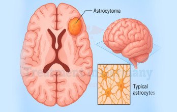
Astrocytomas are gliomas with astrocytic features. In adults, the main diffuse form is astrocytoma, IDH-mutant, meaning the tumor carries a mutation in IDH1 or IDH2. This IDH status is central to the diagnosis and separates these tumors from other diffuse gliomas that can look similar under the microscope, a distinction that matters when planning astrocytoma treatment in Germany.
Older pathology reports often use different terms. Diagnoses made before 2021 may say diffuse astrocytoma, anaplastic astrocytoma, or astrocytoma NOS, labels based on appearance alone under the older classification system. Some files now carry both the old and new names, added after the tissue was retested.
Pilocytic astrocytoma and other pediatric-type astrocytic tumors form a separate category. Pediatric-type diffuse gliomas form separate categories based on their distinct molecular characteristics. They share part of the name but behave very differently from adult diffuse astrocytomas.
Oligodendroglioma
Oligodendroglioma is a diffuse glioma with characteristic oligodendroglial features. It can be difficult to distinguish from astrocytoma by appearance alone. Under current classification, two molecular findings are required for the diagnosis: an IDH mutation and combined deletion of chromosome arms 1p and 19q.
Molecular testing has substantially narrowed this diagnosis by separating tumors that may appear oligodendroglial under the microscope into distinct molecularly defined entities. In one study of 182 tumors previously called oligodendroglioma by microscopy, only 91 met the modern molecular criteria. The older mixed diagnosis oligoastrocytoma has also largely disappeared because molecular testing usually places these tumors into either the astrocytoma or oligodendroglioma group [2].
Glioblastoma
Glioblastoma is the most common malignant primary brain tumor in adults. Doctors now call it glioblastoma, IDH-wildtype, and diagnose it in one of two ways: by how the tumor looks under the microscope, or by specific genetic changes found in the tumor tissue. These include EGFR amplification, a TERT promoter mutation, or the loss and gain of particular chromosomes.
Not every tumor falls clearly into a recognised category. In a sequencing study of 479 tumors, 439 met the criteria for glioblastoma, while the remaining 40 could not be classified into any recognised category [3].
Glioblastoma is only one of many brain tumors. In U.S. data from 2017 to 2021, it made up 13.9% of all primary brain and CNS tumors, but 51.5% of the malignant ones [1].
You may also see the older name, glioblastoma multiforme. Names have changed, and an older diagnosis does not always match today's categories. Tumors once called IDH-mutant glioblastoma are now classified differently, as astrocytoma, IDH-mutant, CNS WHO grade 4. This matters if you are considering glioblastoma treatment in Germany, where molecular testing is routine and may reclassify a tumor diagnosed years earlier.

Glioblastoma as a share of all primary brain and CNS tumors compared with malignant tumors only, United States, 2017 to 2021 [1].
Meningiomas and Other Non-Glial Brain Tumors

Taken together, non-glial tumors account for more primary brain and CNS tumors than gliomas, largely because meningiomas are so common. Their behavior varies widely: some, particularly meningiomas and schwannomas, usually grow outside the brain tissue itself, while others arise within the central nervous system.
Meningioma Tumors
Meningiomas arise from the meninges, the protective membranes around the brain and spinal cord. Most are extra-axial, meaning they grow outside the brain tissue itself and tend to displace rather than diffusely infiltrate it. They are the most common primary CNS tumor and occur more often in women than in men [1].
Meningioma behavior varies considerably. Many grow slowly and are discovered incidentally, while others recur or behave more aggressively. Histologic grade remains important, but molecular features can provide additional information about the risk of progression and recurrence. Location matters as well: a small skull-base meningioma involving important nerves or blood vessels may be more difficult to manage than a larger tumor in a less sensitive position [4].
Other Primary Non-Glial Brain Tumors
The remaining families are smaller, clinically distinct, and each with characteristic anatomical, molecular, and age-related patterns and settings:
- Ependymomas arise from the lining of the ventricles and the central canal of the spinal cord, so they can appear anywhere along that route. Molecular groups now describe them better than location does.
- Schwannomas grow on the sheath around a nerve. Vestibular schwannoma, on the nerve serving hearing and balance, is much the most common, and these are typically non-malignant.
- Pituitary and sellar-region tumors, including pituitary neuroendocrine tumors and craniopharyngiomas. Sitting at the base of the skull, they tend to disturb hormones or vision rather than brain function.
- Pineal-region tumors are uncommon and mixed, running from slow-growing pineocytoma to aggressive pineoblastoma.
- Germ cell tumors trace back to cells that settled in the brain during development. They favor midline sites near the pineal gland or the pituitary stalk, mostly in teenagers and young adults.
- Embryonal tumors such as medulloblastoma are largely a childhood problem, usually arising in the cerebellum, and are now split into molecular groups that shape management.
- Primary CNS lymphoma grows in the brain, the eyes or the spinal fluid, yet it comes from immune cells rather than brain cells. Reduced immune function increases the risk, although many cases also occur in people with normal immune function.
New entities are still being described. Every one of these tumors is uncommon, so experience with the specific type counts for a great deal.
Brain Tumor Type and Molecular Classification

The tumor's type is its diagnostic identity, and it determines which treatments and clinical trials apply to you. Three related terms are often confused with it.
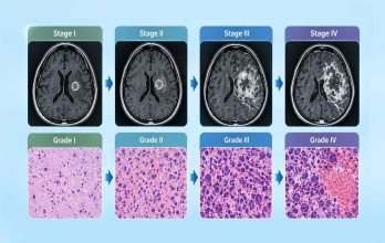
- Grade describes expected behavior, not identity. Under the current WHO system, grade is assigned within a tumor type, so grade 3 in one type doesn't carry the same meaning as grade 3 in another.
- Molecular classification refers to genetic and epigenetic changes in the tumor. These no longer sit alongside the diagnosis as extra information. For many tumors they now define the type itself, and in some cases determine the grade as well.
- Stage, the system used for breast or bowel cancer, isn't applied to most primary brain tumors, because these tumors rarely spread beyond the central nervous system.
Type is established from tissue. A neuropathologist examines the cells, and molecular testing looks for the alterations the classification requires. Where the two disagree, molecular findings often take precedence. This is why a tumor can be renamed while the tumor itself is unchanged.
It also explains why two pathology reports may seem to contradict each other. The WHO classification was revised in 2021, so an older report may use a term since withdrawn. A current diagnosis states the type together with its defining molecular features, as in oligodendroglioma, IDH-mutant and 1p/19q-codeleted, where both features are required for that diagnosis.
The system has limits. DNA methylation profiling supports diagnosis in difficult cases but doesn't replace conventional pathology. Among 1,481 patients in a Hamburg series analyzed with the classifier developed at the German Cancer Research Center, 69 tumors, or 4.6%, matched no known class [5]. That isn't a failed diagnosis. It means one line of evidence was inconclusive, and tumors like these remain treatable by specialists who see them often.
Because the tumor type sets the direction for everything after it, patients considering brain cancer treatment in Germany often start by having their pathology and molecular results reviewed at a specialist center. TIG GmbH (Treatment in Germany) can arrange that review and handle the practical side around it.
Reference


























.webp)
 (1).webp)

.webp)


.jpg)

 (1).webp)


.jpg)