
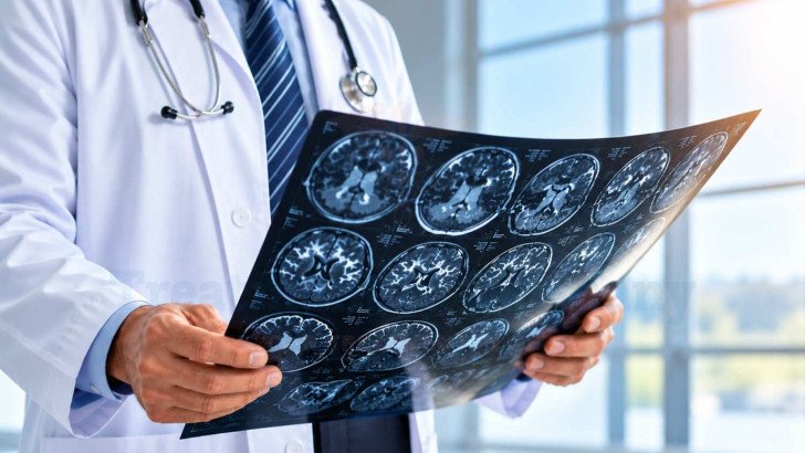
Glioblastoma treatment in Germany involves surgery or biopsy, radiotherapy with temozolomide, and care guided by MGMT status, age, overall health and neurological function. Treatment may also include rehabilitation, follow-up MRI, management of pseudoprogression, options for recurrent disease, selected additional therapies, and multidisciplinary review at specialist neuro-oncology centres.
Glioblastoma Treatment in Germany: Treatment Options, Recovery and Outcomes
Once glioblastoma is confirmed, treatment decisions often need to be made quickly, while patients are still trying to understand the diagnosis and proposed care. The overall treatment pathway is well established, but the exact plan depends on what is safe and appropriate for each patient. In Germany, glioblastoma treatment is planned according to factors such as tumor location, molecular findings, overall health and the extent of surgery that can be safely performed.
Glioblastoma Treatment Planning After Diagnosis
Planning begins with the confirmed pathology and molecular results, since those define the tumor and help determine the appropriate treatment approach. The team then works through the questions that decide what is realistic:
- The tumor's location and extent, its relationship to critical brain structures, and whether maximal safe resection is feasible.
- Current neurological function and performance status.
- Age and general health, including illnesses that affect tolerance of treatment.
- Whether the patient is able and willing to undergo the planned course of radiotherapy and systemic treatment, including the associated recovery and monitoring.
- What the patient themselves wants from treatment.
Glioblastoma usually requires more than one type of treatment because tumor cells can extend beyond the area visible on MRI. For eligible patients, treatment therefore typically combines surgery with postoperative radiotherapy and systemic therapy. When surgery is feasible, it is generally performed first. Radiotherapy and chemotherapy usually follow after recovery, with the treatment plan agreed by a multidisciplinary neuro-oncology tumor board.
Treatment plans vary because age, functional status, tumor biology and how safely the tumor can be removed all affect treatment options. In a German study of 233 patients with newly diagnosed glioblastoma, survival was associated with extent of resection, age, MGMT status and performance status. Clinical trials also tend to include fitter patients, so real-world treatment may differ from published trial protocols [2].
Surgery as Initial Local Treatment for Glioblastoma

Surgery can help confirm the diagnosis by providing tumor tissue for testing and, when possible, remove as much of the tumor as safely possible. The extent of removal depends largely on the tumor’s location and its relationship to important areas of the brain.
Maximal Safe Tumor Removal
When tumor removal is feasible, the aim is maximal safe resection: removing as much tumor as possible while minimizing the risk of new permanent neurological deficits. Greater tumor removal may improve disease control, but preserving functions such as speech, movement and cognition remains the priority.
Complete removal of the tumor visible on MRI is possible in some patients but not in others, especially when the tumor is close to areas controlling important functions or major blood vessels. Even after apparently complete resection, glioblastoma cells can remain beyond the area visible on MRI. This is why additional treatment is needed after surgery.
Biopsy When Tumor Removal Is Not Feasible
For some patients the safest surgical objective is tissue alone. Biopsy is chosen when the tumor lies deep, sits in regions controlling speech or movement, appears in more than one place, or the patient is not well enough for a long operation.
Biopsy is an appropriate option when removing the tumor would carry too much risk. In the German cohort above, 73% of patients underwent resection and 27% had biopsy only. In a Munich series of 1,214 stereotactic biopsies performed for newly diagnosed or suspected recurrent tumors, IDH-wildtype glioblastoma was the most common diagnosis, reported in 596 cases. A biopsy provides tissue for diagnosis and molecular testing but does not significantly reduce tumor volume [2][3].
Radiation and Temozolomide After Glioblastoma Surgery
Once the patient has recovered from surgery, standard postoperative treatment begins. For suitable patients, this usually includes radiotherapy with temozolomide followed by maintenance temozolomide.
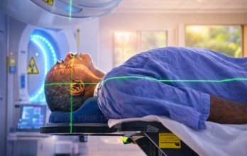
External-beam radiotherapy is usually given over about six weeks, targeting the tumor bed and surrounding areas where infiltrating cells may remain. Temozolomide, an oral chemotherapy, is taken during radiotherapy and then in monthly maintenance cycles. European guidance supports this approach for adults in good general and neurological condition [1].
For older or frailer patients, treatment is adjusted according to general health, neurological function, MGMT status and expected treatment tolerance. Options may include:
- A shorter course of radiotherapy, such as 40 Gy in 15 fractions.
- Short-course radiotherapy with temozolomide in suitable patients.
- Temozolomide alone for selected patients with MGMT-promoter-methylated tumors when combined treatment is not appropriate.
- Radiotherapy alone when chemotherapy is unlikely to provide sufficient benefit or cannot be tolerated.
- Best supportive or palliative care when tumor-directed treatment is unlikely to provide sufficient benefit relative to its potential burdens, based on the patient's overall condition, prognosis, and goals of care.
These are established treatment approaches selected according to the individual patient's condition.
Molecular Factors Affecting Glioblastoma Treatment
MGMT promoter methylation is an important biomarker in glioblastoma. When the MGMT promoter is methylated, the tumor usually produces less of the MGMT DNA-repair enzyme. This makes tumor cells less able to repair the DNA damage caused by temozolomide, so these tumors generally respond better to the treatment.
MGMT status provides both prognostic information about the likely course of the disease and predictive information about potential benefit from temozolomide. However, it does not determine treatment on its own. An unmethylated tumor does not automatically rule out temozolomide, particularly in an otherwise fit patient [1].
IDH status belongs to the diagnosis rather than the plan: glioblastoma is by definition IDH-wildtype, and a grade 4 tumor with an IDH mutation is a different disease. Otherwise, treatment is set by clinical condition, tumor location, imaging and tolerance.
Managing Treatment Effects During Glioblastoma Care

Active treatment can continue for several months, and how a patient feels during this time depends not only on the cancer treatment but also on the supportive care they receive.
Symptoms and Treatment Effects
Fatigue is common during chemoradiation and may increase as radiotherapy progresses. It can also continue for several weeks after treatment ends.
Other possible treatment effects include:
- Nausea and reduced blood-cell count from temozolomide, which is why regular blood tests are required.
- Hair loss and skin changes in the area receiving radiotherapy.
- Neurological symptoms caused by the tumor or treatment-related swelling.
- Seizures, which may require anti-seizure medication or dose adjustment.
- Changes in memory, concentration or word-finding.
Corticosteroids such as dexamethasone may be used to reduce brain swelling and related symptoms. Because longer use can cause significant side effects, the dose is usually reduced when clinically possible.
Rehabilitation and Daily Function
Rehabilitation can begin during active treatment and is tailored to the patient's symptoms and level of independence. It may include:
- Physiotherapy for strength, balance and mobility.
- Occupational therapy for daily activities and adaptations at home.
- Speech and language therapy when communication or swallowing is affected.
- Cognitive rehabilitation for difficulties with memory, attention and planning.
Needs vary and change over time. In a prospective study of 92 patients needing prolonged inpatient rehabilitation after primary brain tumor surgery, those with malignant tumors started from a poorer functional baseline yet required comparable input [5].
Monitoring Glioblastoma and Follow-Up Imaging
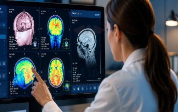
Follow-up combines the patient's symptoms and neurological examination with MRI findings. An early postoperative MRI is usually performed within 24 to 48 hours after surgery to assess residual tumor. Further MRI scans are commonly performed every few months, although the timing may be adjusted according to symptoms and the stage of treatment. Increased enhancement on MRI does not always mean that the tumor is growing.

Three different processes that can look similar on MRI. Pseudoprogression and radiation necrosis are not the same thing and should not be used interchangeably.
Pseudoprogression is most important to consider during the first few months after chemoradiation. In a study of 169 patients whose MRI showed early radiographic progression, 38.5% met the study's definition of clinically defined pseudoprogression, and it was more common in MGMT-promoter-methylated tumors [4].

Classification of 169 glioblastoma patients with early radiographic progression after chemoradiation [4].
Several factors help doctors distinguish treatment effects from tumor progression:
- How long it has been since radiotherapy ended.
- Whether neurological symptoms are stable or worsening.
- Which treatments have recently been given.
- How the abnormality changes on repeat MRI scans.
When uncertainty remains, advanced MRI techniques, amino-acid PET or, occasionally, a tissue sample may provide additional information. A single concerning MRI does not by itself confirm that treatment has failed.
Treatment After Glioblastoma Progression or Recurrence
Glioblastoma recurs in most patients. When this happens, the treatment plan is reassessed based on where the tumor has returned, previous treatment, the patient's general condition and treatment goals.
Options may include:
- Repeat surgery when the recurrent tumor can be removed safely.
- Reirradiation in selected patients, depending on the previous radiation dose and treatment interval.
- Further systemic treatment based on treatments already received.
- Participation in an appropriate clinical trial.
- Supportive or palliative care when further tumor-directed treatment is unlikely to provide sufficient benefit.
At recurrence, surgery still aims to remove as much tumor as safely possible while preserving neurological function. In the RANO resect study, more extensive removal of non-enhancing tissue was not associated with better survival and was linked with a greater risk of new neurological deficits [6].
There is no single treatment that is appropriate for every recurrence. The decision depends on the location of the tumor, previous treatments, the time since initial treatment and the patient's condition and preferences.
Additional and Emerging Treatment Approaches for Glioblastoma
Several additional treatments may be discussed with patients, but their evidence, indications and availability differ. Some are used in selected patients, while others remain investigational.
- Tumor Treating Fields (TTFields): low-intensity alternating electric fields delivered through arrays placed on the scalp. In Germany, TTFields may be considered in eligible patients after chemoradiation as part of maintenance treatment.
- Carmustine wafers: chemotherapy implants placed in the surgical cavity during surgery. They are used selectively rather than routinely particularly in patients undergoing resection, but their role in the era of contemporary radiotherapy and temozolomide remains uncertain, and they are not routinely used in all patients.
- Bevacizumab: an antiangiogenic treatment that may help control edema and symptoms in selected patients with recurrent disease, but is not standard first-line treatment.
- Immunotherapy, vaccines and cell therapies: currently investigational for glioblastoma and generally used within clinical trials.
Evidence, regulatory approval and availability are separate considerations. A treatment described as promising may still be investigational and may not yet be part of standard glioblastoma care.
Glioblastoma Treatment and Consultation in Germany

Glioblastoma treatment in Germany is available at university hospitals and specialist cancer centres with multidisciplinary teams. Neurosurgery, neuro-oncology, radiation oncology, neuropathology and neuroradiology can work together to assess complex cases and plan treatment. Certified cancer centres follow defined quality standards for multidisciplinary care, and the WiZen study found an association between treatment at certified hospitals and lower mortality, although the neuro-oncology subgroup result was not statistically significant after adjustment for multiple comparisons [7].
A specialist review can be useful at diagnosis, when imaging is unclear, or when the tumor progresses. Patients can have their pathology, molecular results and MRI scans reviewed by German specialists before deciding on further treatment.
TIG GmbH (Treatment in Germany) helps international patients arrange specialist reviews and complete logistical arrangements, including travel, accommodation and medical visa support where required, for those seeking brain cancer treatment in Germany.
References
Why Patients Worldwide Prefer Our Medical Services in Germany – Key Benefits Explained













.webp)
 (1).webp)
.webp)

.jpg)
 (1).webp)
.jpg)