
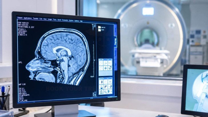
Brain tumor diagnosis combines clinical assessment, imaging, tissue analysis and molecular testing to determine what a brain lesion is and how it should be classified. MRI, CT and advanced imaging help define the lesion, while biopsy, histopathology and molecular tests can confirm the diagnosis. It also explains why diagnoses may change, when second opinions or further review may be needed, and how specialist brain tumor diagnosis is organized in Germany.
Brain Cancer Diagnosis: MRI, Biopsy and Pathology
An abnormal brain scan starts the diagnostic process but does not confirm a brain tumor. Imaging can locate a lesion and suggest likely causes, while tissue analysis, when needed, can identify the tumor type and molecular changes. These findings help doctors determine the most appropriate brain cancer treatment in Germany for each patient.
Initial Assessment for a Suspected Brain Tumor
Investigation usually starts for one of three reasons: new neurological symptoms, an abnormal finding on examination, or a lesion noticed by chance on a scan done for something unrelated, such as a head injury. The first assessment brings together what is already known:
- Medical history, including when problems began and how they have changed, earlier cancers, other illnesses and current medication.
- Neurological examination of strength, sensation, reflexes, coordination, vision, speech and thinking, which shows whether the lesion is affecting function.
- Previous records, particularly older brain scans, because comparison over time can help determine whether a lesion is stable, newly developed, or changing in size or appearance.
- Review of existing imaging by a neuroradiologist, who judges whether the available scans are adequate or a dedicated tumor protocol is needed.
Until the evidence is complete, the accurate term is a suspected lesion. The word “tumor” in a scan report reflects a radiological impression, and infection, inflammation, demyelination and stroke can all produce a similar picture. A brain tumor diagnosis is confirmed, not assumed.
Brain Imaging for Tumor Diagnosis

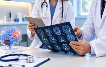
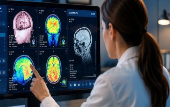
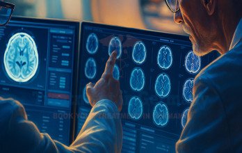
MRI is the main imaging test. It shows brain tissue in greater detail than CT and does not use ionizing radiation although CT remains particularly useful for acute hemorrhage, calcification, bone abnormalities, and situations requiring rapid imaging. A tumor protocol combines several sequences, often including gadolinium-enhanced images that highlight areas where the blood-brain barrier is disrupted. Together, the images describe:
- The position and size of the lesion, and whether there is more than one.
- Whether it arises within brain tissue or from surrounding structures such as the meninges.
- Its internal pattern, including contrast uptake, cystic or necrotic components, hemorrhage, and other structural features.
- Swelling around it and pressure on neighboring structures or fluid spaces.
Specialized techniques are added when the standard scan leaves questions open. Perfusion MRI estimates blood flow within the lesion, diffusion imaging reflects how tightly cells are packed, and MR spectroscopy measures chemical signals in the tissue. Amino acid PET highlights metabolically active tumor and can point to the most informative area to sample.
Even the best imaging describes a lesion but it does not always establish the exact tumor type or molecular diagnosis. Different tumor types can look alike, and some non-tumor conditions imitate tumors closely. That is why a good MRI report lists the likely possibilities instead of a single answer with relevant alternative possibilities and recommendations for further imaging or tissue diagnosis when the findings are not specific.
Biopsy and Tissue Diagnosis for Brain Cancer

When imaging leaves the diagnosis open, tissue sampling is often required for definitive diagnosis and molecular characterization. Tissue analysis can establish what a scan only suggests: whether the lesion is a tumor at all, and what kind.
The decision to obtain tissue is made case by case. Doctors weigh how much the result would change management, the risk of reaching the lesion, and whether it can be removed at the same time. A deep lesion close to areas controlling speech or movement is approached differently from one near the surface although location alone does not determine whether biopsy or resection is appropriate. For a small number of lesions with very typical imaging, such as many meningiomas, clinicians may initially use MRI surveillance rather than immediate tissue sampling when the clinical and radiological findings support observation. Tissue can come from two routes:
- Stereotactic biopsy: a needle is guided through a small opening in the skull to a target planned on imaging. It is used mainly for deep, multiple or hard-to-reach lesions, or when a diagnosis is needed before larger surgery is considered.
- Tissue removed during an operation: when a lesion is accessible, it can be removed partly or fully, which provides more material and combines diagnosis with removal.
Stereotactic biopsy has a high diagnostic yield although results vary according to lesion characteristics, sampling technique, the suspected diagnosis, and institutional experience. In a Munich series of 1,214 procedures for suspected glioma, 96.3% produced an integrated diagnosis. In a US series, 23 of 387 biopsies (5.94%) were non-diagnostic, and 60.8% of those were later classified as false negatives. An inconclusive biopsy does not mean the lesion is harmless and further clinical, radiological, pathological, or, in selected cases, repeat tissue assessment may be required [1] [2].
Pathology and Molecular Testing for Brain Cancer Diagnosis
Once tissue reaches the laboratory, neuropathology examines it in layers, and each layer adds information the previous one cannot provide:
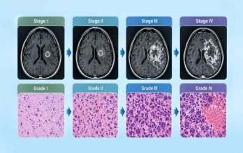
- Histopathology: stained sections are examined under the microscope for cell type, arrangement, density, including cell morphology, cellularity, mitotic activity, necrosis, microvascular proliferation, and other features relevant to tumor classification and grading.
- Immunohistochemistry: antibodies bind to specific proteins, showing the cells’ origin and, for some key mutations, detecting them directly
- Molecular testing: DNA and sometimes RNA are analyzed for mutations, chromosomal changes and gene fusions, often with sequencing panels
- DNA methylation profiling: the tumor’s pattern of chemical tags on its DNA is compared with a reference library of known tumor classes
In a Belgian series of 36 adults with mainly difficult diagnoses, methylation profiling confirmed the diagnosis in 33%, refined it in 11% and changed it in 8%; 39% produced only a low-confidence match. These figures should not be generalized to all brain tumors [3].

Effect of DNA methylation profiling on the diagnosis in 36 adults with challenging brain tumors. Source: Lebrun et al [3].
Confirming and Clarifying a Brain Cancer Diagnosis
The final diagnosis combines pathology and molecular findings with the clinical and imaging picture. If the results conflict, specialist review or further testing may be needed.

An early imaging impression can differ from the final result for several reasons. Two tumor types may look alike on MRI, a lymphoma or an infection may resemble a glioma, or a biopsy may capture a less representative part of the lesion. Some tumors are simply unusual and fit no established category well. When findings conflict or remain uncertain, the usual next steps are:
- Review of the slides by a second neuropathologist, often at a reference center.
- Additional immunohistochemistry or molecular testing, including methylation profiling.
- correlation of tissue and imaging, to confirm that the sampled area matches the suspicious region.
- Discussion at a multidisciplinary tumor board, where neuroradiology, neuropathology, neurosurgery and neuro-oncology review all results together.
- Occasionally a repeat biopsy or short-interval follow-up imaging.
A diagnosis may change when more complete molecular information becomes available. In a German study of more than 1,200 children and adolescents, neuropathology and methylation-based classification disagreed in 30% of cases and affected clinical management in 5% [4]. These figures should not be applied directly to adults.
Specialist review or a second opinion is most useful when the tumor is rare, when the report describes it as “not otherwise specified”, when imaging and tissue findings do not match, or when the diagnosis will drive a major decision. It works best with the original MRI files and the pathology material itself, not only the written reports.
Specialist Diagnosis for Brain Tumors in Germany

Diagnosing a brain tumor usually involves several specialists. Neurologists and neurosurgeons assess the patient, neuroradiologists review the imaging, and neuropathologists examine tumor tissue and molecular findings. In specialist German centers, complex cases may also be discussed at a multidisciplinary tumor board.
For difficult or unusual cases, additional diagnostic methods may help:
- Methylation testing: Can help classify brain tumors more precisely when the diagnosis is unclear.
- Second-opinion pathology: Specialist neuropathologists can review tissue again when confirmation is needed.
- Image-guided biopsy planning: In selected cases, PET and MRI may be combined to help identify the most informative area for biopsy.
Not every center performs every test on site, so some samples may be sent to a specialist laboratory. A second opinion can often begin with existing imaging, pathology, molecular results and tissue material, allowing specialists to decide whether the diagnosis is confirmed or whether further testing is needed.
TIG GmbH (Treatment in Germany) coordinate this process for patients seeking specialist brain tumor diagnosis or a second opinion in Germany. This can include collecting and transferring medical records, imaging and pathology material, communicating with specialist hospitals and laboratories, arranging medical reviews and appointments, and coordinating the practical steps needed if further testing or treatment is recommended.
References













.webp)
 (1).webp)
.webp)
.jpg)
 (1).webp)
.jpg)