
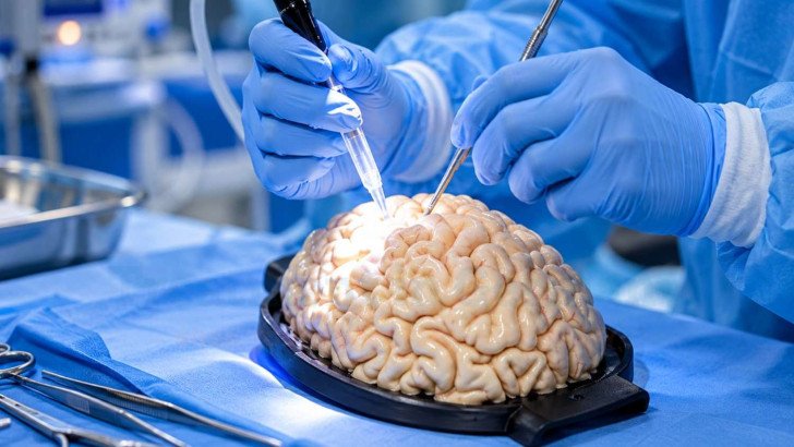
Brain tumor surgery may involve stereotactic biopsy, maximum safe resection or debulking, depending on the goal of treatment. Surgical planning also considers techniques that help protect speech and movement, including neuronavigation, functional mapping and awake craniotomy. Recovery includes early postoperative care, surgical risks and rehabilitation, while pathology and postoperative imaging help show what was removed and guide further treatment planning.
Brain Cancer Surgery in Germany: Tumor Removal, Recovery and Specialist Care
Brain tumor surgery does not always aim for complete removal. The surgical goal depends on the tumor’s type, location and the individual patient. Surgery may be used to obtain tissue for diagnosis, relieve pressure caused by the tumor, or remove as much of the tumor as possible while preserving important functions such as speech and movement. This is also an important part of planning brain cancer treatment in Germany.
Surgical Decision Making for Brain Tumors
The surgical plan depends on several factors:
- Tumor location and proximity to areas controlling involved in language, motor, sensory, visual, cognitive, or other critical functions.
- Tumor size, boundaries and pattern of growth.
- Whether the tumor can be reached without damaging critical brain tissue and resected while preserving critical neurological functions.
- Pre-existing neurological, cognitive, language, or functional deficits and the patient’s baseline functional status.
- Overall medical condition, comorbidities, functional status general health and fitness for anaesthesia.
- The patient’s priorities when balancing greater removal against neurological risk.
A stereotactic biopsy may be chosen instead of tumor resection when a tissue diagnosis is needed but removal would be unsafe or is not expected to provide sufficient benefit. This may apply to deep-seated lesions, tumors close to critical brain structures, selected patients with multiple lesions, or patients whose overall condition makes a longer resection unsuitable.
This is the principle of maximum safe resection:
removing as much tumor as possible while minimizing the risk of permanent neurological or functional impairment. The intended extent of surgery is planned beforehand, often with multidisciplinary input, and should be discussed with the patient before consent.
Brain Tumor Biopsy and Surgical Removal

The type of brain tumor surgery depends on its purpose. Surgery may involve obtaining tissue for diagnosis, removing as much tumor as safely possible, or intentionally removing only part of the tumor.
Biopsy for Tissue Diagnosis
When tissue is needed but resection is not appropriate, stereotactic biopsy uses MRI-based navigation to guide a needle through a small opening in the skull and collect tissue samples.
In a series of 1,214 stereotactic biopsies, 95.9% caused no clinical sequelae, while severe or life-threatening complications occurred in 1.2%; no procedure-related deaths were reported. Bleeding is an important procedural risk [1].

Clinical outcomes across 1,214 consecutive stereotactic biopsies for suspected glioma [1]. Most procedures caused no symptoms at all.
Resection also provides tissue for diagnosis, so a separate biopsy is usually unnecessary. Biopsy establishes the diagnosis but does not reduce tumor volume.
Maximum Safe Tumor Removal
When resection is planned, surgeons remove as much tumor as possible without causing unacceptable neurological harm and and avoiding any permanent neurological deficits.
For glioblastoma, RANO criteria classify surgery as supramaximal, maximal or submaximal resection, or biopsy alone. A population-based study of 470 patients validated these categories although the survival differences between categories were smaller than those reported in the original study. Patients may hear simpler terms such as complete, near-complete or partial removal [2].
Complete removal may be possible for well-defined tumors such as many meningiomas and metastases although microscopic tumor cells may remain beyond the apparent surgical margin. Diffuse gliomas infiltrate surrounding brain, so tumor cells may remain even after all visible tumor is removed. Tumor may deliberately be left when:
- It lies within tissue controlling speech or movement, through preoperative assessment and, when indicated, intraoperative functional mapping.
- It is wrapped around a major artery or vein that cannot be sacrificed.
- It sits against the brainstem, where there is no safe margin.
- The patient’s condition means the operation has to be brought to a close.
Debulking When Complete Removal Is Not Possible
Debulking is intentional partial removal when complete resection would be unsafe or would carry an unacceptable risk of neurological or other serious complications. It aims to reduce tumor volume, pressure or symptoms while preserving neurological function. Patients should understand that residual tumor is expected.
Techniques for Protecting Brain Function During Surgery

Several surgical technologies help neurosurgeons locate the tumor, identify critical brain areas and reduce the risk of neurological injury during resection. These techniques do not eliminate surgical risk but can support safer and more precise tumor removal and help balance tumor removal with preservation of neurological function.
Neuronavigation and Image-Guided Surgery
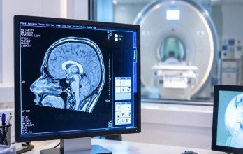
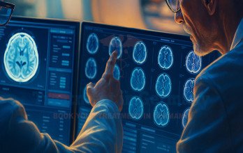
Neuronavigation links preoperative MRI scans to the patient’s head position, helping surgeons locate the tumor and avoid critical structures during surgery.
Brain shift during surgery can reduce the accuracy of preoperative navigation because the relationship between the tumor and surrounding structures may change during the procedure. Intraoperative ultrasound or MRI and, in selected gliomas, fluorescence-guided surgery can help compensate and identify tumor tissue that may otherwise be difficult to distinguish from surrounding brain [3].
Functional Mapping, Monitoring and Awake Surgery
Navigation shows anatomy, while functional mapping identifies brain regions responsible for functions such as movement or language by electrically stimulating selected areas during surgery.
Motor pathways can be monitored under general anaesthesia, but language testing requires a responsive patient. Awake craniotomy may therefore be used when surgery involves language-related areas.
During awake craniotomy, anaesthesia or sedation is used for painful parts of the procedure, while the patient remains responsive during functional testing. Tasks may include naming pictures, speaking or moving a limb while the surgeon maps critical brain areas.
In a study of 80 patients undergoing awake mapping for gliomas in eloquent areas, 17.5% had neurological deficits lasting less than a week and 15% had deficits at three months; 90% had a Karnofsky Performance Status above 80 at three months [4].
Assessment and Preparation Before Brain Tumor Surgery

Preoperative assessment helps the surgical team plan the procedure safely and document the patient’s neurological function before surgery. This baseline can also help clinicians assess changes during recovery.
A typical workup includes:
- Recent MRI, including navigation sequences when needed, and review of existing pathology.
- Aneurological examination recording strength, sensation, coordination, vision and speech.
- Targeted language, cognitive or visual testing when relevant.
- Blood tests, heart and lung assessment, and a medication review, especially anything affecting clotting and antiplatelet drugs, and other medications that may affect bleeding or anesthesia.
- An anaesthetic consultation, which before an awake procedure includes rehearsing the tasks used in theatre.
Questions to ask the surgeon include:
- What is the goal of this operation, and how much removal is realistically expected?
- Which functions are at risk, and what would a bad outcome look like?
- What happens if the tumor proves more extensive than the scan suggested?
- How long is the hospital stay likely to be, and is rehabilitation expected?
- When will the pathology result be available, and who will explain it?
Recovery, Risks and Findings After Brain Tumor Surgery
Recovery after brain tumor surgery varies between patients and depends on the tumor, surgical procedure and neurological function before surgery. Physical healing and neurological functions such as strength, speech, coordination, cognition, and vision may progress at different rates.
Early Recovery After Brain Tumor Surgery
After surgery, patients are closely monitored in a recovery, neurosurgical or intensive care unit. Staff regularly check consciousness, pupils, strength and speech for early signs of complications. The first days may involve:
- Early postoperative MRI when indicated, often within 24–48 hours after glioma resection.
- Headache, fatigue and swelling managed with medication.
- Temporary worsening from postoperative swelling.
- Early mobilisation, sometimes with physiotherapy.
- Discharge after several days if recovery is uncomplicated.
Fatigue may persist for several weeks even after uncomplicated surgery. Return to driving, flying and work is decided individually.
Neurological Effects and Surgical Risks
Surgical risk varies with tumor location, extent of resection and the patient’s condition before surgery.

The principal risks of brain tumor surgery. Individual likelihood varies widely with tumor site and the extent of removal.
A new neurological deficit after surgery is not always permanent. Some deficits improve as postoperative swelling and temporary tissue disturbance resolve.
Rehabilitation and Functional Recovery
Rehabilitation may begin soon after surgery and continue according to the patient’s neurological and functional needs.
- Physiotherapy for strength, balance and walking.
- Occupational therapy for everyday activities.
- Speech and language therapy for communication and swallowing.
- Neuropsychological rehabilitation for memory, attention and planning.
In a study of 92 patients requiring prolonged rehabilitation after primary brain tumor surgery, those with malignant tumors had poorer functional status, but malignant and non-malignant groups had similar rehabilitation needs [5].
Pathology and Postoperative Findings
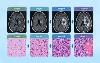
Removed tissue is examined by neuropathology using microscopy and, when appropriate, molecular testing. Resection generally provides more tissue than needle biopsy for diagnosis.
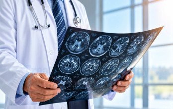
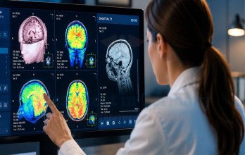
Postoperative MRI helps assess residual tumor and provides a baseline for later scans. Imaging and pathology findings then guide further treatment planning.
Brain Tumor Surgery and Specialist Care in Germany

Brain tumor surgery in Germany is performed mainly at university hospitals and larger neurosurgical departments, and the services available differ between them. Tumors that are difficult to reach, located near the brainstem, or close to the areas of the brain that control speech and movement are usually treated at centres that manage such cases regularly.
What distinguishes a specialist centre is not only its equipment but how the specialties work together. Neurosurgery, neuro-oncology, neuroradiology, neuropathology and radiation oncology review the case at a joint tumor board, so the aim of the operation is agreed together with the treatment that will follow.
Surgical technology is widely available but not everywhere. Nationwide data covering 101,192 malignant glioma cases in Germany between 2019 and 2022 showed that, among operations to remove tumor, microsurgical technique was used in 95% and intraoperative navigation in 84% [6]. Functional mapping and monitoring during surgery are offered by some departments and not others. The German Cancer Society (Deutsche Krebsgesellschaft) certifies neuro-oncology centres against standards for case numbers, interdisciplinary review and guideline-based care, which is a helpful guide rather than a guarantee.
Much can be clarified before travelling. Patients considering brain cancer treatment in Germany can have their existing MRI scans and original pathology reports reviewed remotely, allowing doctors to assess the diagnosis and whether surgery may be an option before the patient travels.
Questions worth asking any centre:
- How experienced is the surgical team with tumors in this location?
- Which surgical techniques may be suitable for my tumor, and are these available at the centre?
- What is the goal of surgery in my case: taking a biopsy, removing part of the tumor, or removing as much as safely possible?
- Will my case be reviewed by a specialist neuro-oncology team before surgery?
- What should I expect during recovery, and how long might I need to stay in hospital?
Patients can have their MRI scans and pathology reports reviewed by German neurosurgical teams before deciding on an operation, including whether removal is feasible and what the realistic surgical goal would be. TIG GmbH (Treatment in Germany) helps international patients arrange this medical review and provides complete logistical support, including travel and medical visa arrangements where required.
References













.webp)
 (1).webp)
.webp)
.jpg)
 (1).webp)
.jpg)