
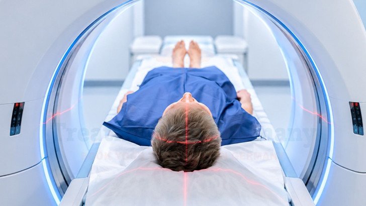
Proton therapy can reduce radiation exposure to healthy tissue in selected patients with brain tumors, but its advantage over modern photon radiotherapy varies between individuals. Suitability depends on tumor location, treatment volume, nearby critical structures and previous treatment. Specialist assessment in Germany may include imaging review and comparison of proton and photon treatment plans.
Proton Therapy for Brain Tumors in Germany: Patient Selection, Planning and Outcomes
Proton therapy for brain tumors in Germany is considered when radiotherapy is indicated and a proton plan may reduce radiation exposure to healthy brain tissue or critical structures compared with a well-optimised photon plan. The expected benefit depends on the tumor’s location, treatment volume and surrounding anatomy, so suitability is assessed individually.
Proton Therapy Role and Patient Selection for Brain Tumors
Proton therapy is a form of external-beam radiotherapy used when radiation is already indicated with modality selection based on tumor characteristics, treatment goals, and the expected dose distribution to the target and surrounding normal tissues. Protons release most of their energy near a planned depth and then undergo a rapid dose fall-off, resulting in little or no exit dose beyond the target. Photons, by comparison, continue depositing radiation as they pass beyond the target although modern techniques such as IMRT and VMAT can substantially reduce and shape the dose delivered to surrounding normal tissues.
Reducing radiation exposure beyond the target is most relevant when nearby healthy tissues or critical structures need to be protected, particularly in patients expected to live long enough for late effects to develop.
Important structures may include:
- The optic nerves and chiasm, involved in vision.
- The brainstem, which controls vital neurological functions.
- The cochlea, involved in hearing.
- The hippocampus, involved in memory.
- The pituitary gland and hypothalamus, which regulate hormones.
- Healthy brain tissue outside the treatment target.
Specialists weigh several things together:
- Tumor type and location, and how close it sits to structures that must be protected.
- Target size and shape, since the advantage varies with the volume being treated.
- Age and expected long-term survival, because late effects take years to surface.
- Previous radiation, including dose and field, where a further course is being considered.
- General health and treatment goals, which shape how much any difference in dose matters.
A diagnosis alone does not determine suitability for proton therapy. Selection often involves comparing a proton plan with an appropriate photon plan for the same patient. In a Danish study of adults with IDH-mutant grade 2 and 3 glioma, age and clinical target volume were associated with proton selection in univariable analysis; clinical target volume remained significant after multivariable adjustment [1].
Proton Therapy Compared With Photon Radiation
Modern photon techniques such as IMRT and VMAT can shape the high-dose region closely around the target while reducing radiation exposure to nearby healthy structures. Proton therapy may further reduce low- and intermediate-dose exposure in selected patients, but the size of this advantage depends on the target location, shape, beam arrangement and relationship to nearby critical structures. It is also important to distinguish dosimetric benefit from clinical benefit.
Lower radiation exposure to healthy tissue does not automatically mean fewer symptoms, better quality of life or improved tumor control. Clinical outcomes need to be demonstrated in patient studies.

In 23 adults with brain tumors, proton plans reduced dose to the left temporal lobe, but the estimated reduction in risk of verbal-fluency decline was modest [2]. These are modelled estimates, not measured outcomes.
Proton Therapy Treatment Planning, Delivery and Course
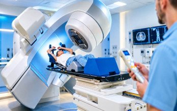
The treatment experience is broadly similar to photon radiotherapy, but proton therapy requires additional planning to account for beam range and anatomical uncertainty.
After specialist assessment and review of MRI, CT and any previous treatment, the process runs through several stages:
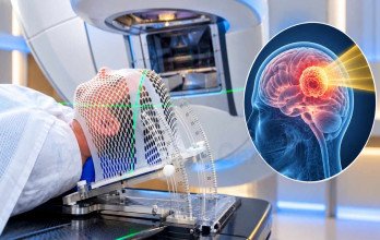
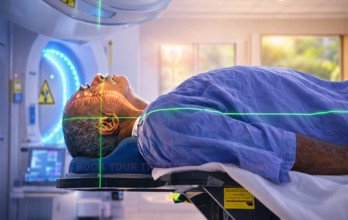
- Simulation, a planning scan in the exact treatment position, with a thermoplastic mask for immobilization, commonly a customized thermoplastic mask for cranial treatments.
- Contouring, where the target and organs at risk are outlined on the fused imaging.
- Plan building, in which a physicist creates the proton plan, usually alongside a photon plan with a comparative modern photon plan generated when clinically appropriate.
- Accounting for range uncertainty: because protons stop at a depth that depends on the tissue they cross, plans are made robust to small shifts in density or position, and beams are arranged so a proton’s stopping point does not fall directly on a critical structure where possible.
- Quality assurance of the plan before treatment starts, and image guidance to verify position at every session.
Courses often mirror the equivalent photon schedule, commonly daily sessions on weekdays over several weeks, though the exact number follows the diagnosis, the target and the plan.

Proton Therapy for Different Brain Tumors
Proton therapy may be particularly relevant when reducing dose to nearby healthy tissue is clinically important, especially in younger patients and those expected to survive for many years. Tumor type alone, however, does not determine whether protons provide a meaningful advantage.
- Selected lower-grade gliomas, where long survival makes late effects on memory and thinking a real concern.
- Meningiomas and other skull-base tumors, which often sit beside the optic apparatus, brainstem and cranial nerves.
- Ependymomas, particularly in the posterior fossa near the brainstem in which the target is close to the brainstem or other critical structures.
- Craniopharyngiomas and pituitary or sellar-region tumors, where the hypothalamus, pituitary and optic chiasm are nearby.
- Many childhood brain tumors, where reducing radiation exposure to healthy developing tissues may be particularly important.
Clinical benefit varies by tumor and anatomy. In the German KRANIOPHARYNGEOM 2007 study, five-year event-free survival in children with craniopharyngioma was similar after proton and photon radiotherapy (92% versus 91%), and radiation doses to the hypothalamus and pituitary were also similar. When a critical structure directly overlaps or closely abuts the treatment target, proton therapy may have limited ability to reduce its dose [4]. Recurrent tumors raise a different question, because the surrounding tissue has already absorbed a first course. Proton reirradiation is considered in carefully assessed patients, and the decision turns on the earlier dose and field, the time elapsed, the current tumor position and how the anatomy has changed since. Specialist review of the original radiation records is essential.
Proton Therapy Side Effects, Recovery and Long-Term Considerations

Proton and photon radiotherapy can cause similar types of acute and long-term side effects. The individual risk depends largely on the radiation dose, the volume treated and which brain structures receive radiation. Proton therapy may reduce exposure to some healthy tissue, but it does not eliminate toxicity.
During and shortly after treatment, patients may experience:
- Fatigue, which usually builds gradually through the course.
- Headache and nausea, often related to swelling in the treated area.
- Hair loss and scalp changes where the beams enter.
- Temporary worsening of neurological symptoms, caused by inflammation or swelling.
Long-term effects depend mainly on which structures receive radiation and the dose they receive:
- Changes in memory, attention or processing speed.
- Hormone deficiencies if the pituitary gland or hypothalamus receives clinically relevant radiation exposure.
- Radiation necrosis within or near the treated area.
Proton therapy does not eliminate long-term treatment risks. In the German KiProReg registry of 294 children treated with proton therapy for central nervous system tumors, 17 developed grade 3 CNS adverse events during long-term follow-up. For adults treated with photon or proton radiotherapy for brain and skull-base tumors, European Particle Therapy Network guidance recommends structured long-term assessment of neurological, neurocognitive and endocrine outcomes [3] [5].
Proton Therapy Evidence, Outcomes and Limitations
Evidence is strongest for reduced radiation exposure to healthy tissue. Evidence that this consistently translates into better long-term clinical outcomes remains more limited and varies by tumor type and patient population.
Type of benefit | What it means | How well established |
|---|---|---|
Dosimetric benefit | Lower radiation dose to selected healthy tissue on the treatment plan | Consistently shown in planning studies for many brain tumors |
Modelled clinical benefit | Predicted reduction in side effects, estimated from dose-effect models | Often favourable but modest; depends on the model used [2] |
Measured clinical benefit | Observed improvement in toxicity, quality of life or disease control | Limited, largely observational, and varies by tumor and population |
Much of the clinical evidence comes from registries and observational studies rather than randomised trials. In the KiProReg pediatric cohort, three-year local control was 79.5%, but the study included different central nervous system tumor types and this figure should not be interpreted as evidence that proton therapy improves tumor control [5].
Evidence for proton reirradiation remains limited. In a prospective multi-institutional registry of 143 adults with recurrent malignant glioma, radiographic radiation necrosis occurred in 19%, while acute and late grade 3 toxicity occurred in 7% and 4%, respectively. In a series of 11 children reirradiated for tumors close to the brainstem, 9 achieved local control and no CNS toxicity of grade 2 or higher was observed. These small and observational studies support feasibility in selected patients but do not establish superiority over photon reirradiation [6] [7].
Selection bias remains an important limitation because proton and photon patient groups may differ in age, tumor location and general health.
Specialist Proton Therapy for Brain Tumors in Germany
Proton therapy in Germany is available at specialist centres, often as part of a wider team that may include radiation oncologists, neuro-oncologists, neurosurgeons and neuroradiologists. Whether it is suitable depends on the individual tumor, previous treatment and whether proton therapy is expected to offer a meaningful advantage over other radiation options.
For patients coming from abroad, the process usually starts with a review of medical records and imaging. If radiation therapy has been given before, the earlier treatment details may also need to be reviewed. A specialist consultation can then help determine whether proton therapy is appropriate before travel is arranged.
How can I find out whether proton therapy is suitable for my brain tumor in Germany?
A specialist can review the tumor type and location, previous treatment and available imaging. When needed, proton and photon treatment plans may also be compared to see whether proton therapy is likely to reduce radiation exposure to healthy tissue in a meaningful way.
For international patients considering brain cancer treatment in Germany, TIG GmbH (Treatment in Germany) can help coordinate the review of medical records and imaging and arrange consultations with specialist radiation oncology teams. The treating medical team remains responsible for deciding whether proton therapy is clinically suitable and for making all treatment decisions.
References








.webp)
 (1).webp)
.webp)
.jpg)
 (1).webp)
.jpg)