
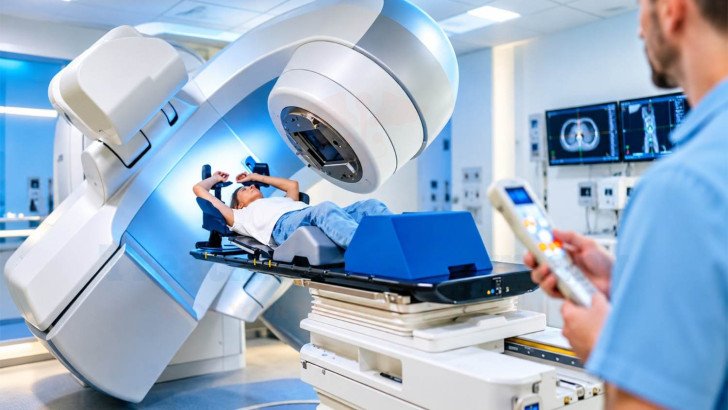
Radiation therapy can be an important part of brain tumor treatment after surgery or for selected tumors that cannot be safely removed. Treatment depends on tumor type, grade, molecular features and previous treatment. This overview covers radiation planning, techniques, treatment schedules, side effects, follow-up imaging and reirradiation, along with how international patients can access specialist radiation care in Germany.
Brain Cancer Radiation Therapy in Germany: Treatment, Planning and Recovery
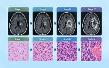
Radiation therapy may still be recommended after a brain tumor has been surgically removed because microscopic tumor cells can remain beyond what is visible on imaging or can be safely removed during surgery. Whether brain cancer radiation therapy in Germany is needed depends on the tumor type, WHO grade, molecular features, extent of resection, previous treatment and the patient’s overall condition.
The Role of Radiation Therapy in Brain Tumor Treatment
Radiation therapy is a local treatment directed at a defined treatment area to a defined target volume while attempting to limit radiation exposure to surrounding healthy tissue. The extent of resection is an important part of treatment planning and can be described using defined categories ranging from complete resection to biopsy alone [2].
Radiation may be considered:
- After surgery for higher-grade tumors, where it commonly forms part of established treatment [1].
- For selected lower-grade tumors with residual or progressive disease.
- For selected meningeal and other CNS tumors.
- For selected brain metastases.
- For symptom relief when long-term disease control is not the primary goal.
Treatment intent influences the radiation dose and schedule. Palliative radiation describes the treatment goal rather than an individual patient’s prognosis. The decision also depends on tumor type, grade, molecular findings, location, residual disease, previous treatment and performance status. In a German study of 233 patients with newly diagnosed glioblastoma, treatment patterns varied with factors including age, extent of surgery and performance status [3].
Planning and Delivering Brain Radiation Therapy

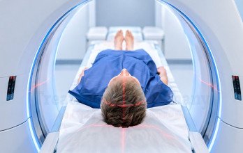
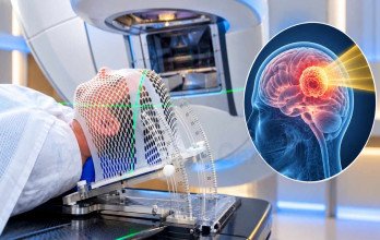
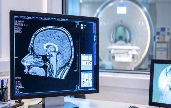
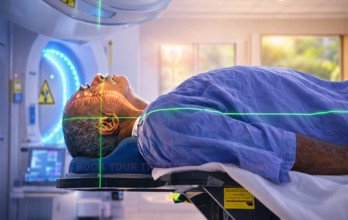
Planning begins with review of imaging, pathology, molecular findings and previous treatment. If radiation is recommended, CT simulation is performed in the position that will be used during treatment. A thermoplastic mask is moulded around the head and face to minimise movement and reproduce the same position at each session. It is close-fitting but should not be painful it should be tolerable, although some patients may experience pressure, anxiety or discomfort while wearing it.
From the planning scan, the team defines what is to be treated and what must be protected:
- The target volume, drawn from the planning CT combined with MRI and the operative findings.
- Organs at risk, including the optic nerves and chiasm, brainstem, cochlea, lens and, where relevant, the hippocampus.
- The dose distribution, planned to treat the target while limiting exposure to surrounding tissue.
- Quality assurance checks on the plan and the machine before any dose is delivered and relevant image-guidance processes before treatment, with additional safety checks performed according to the center's protocol.
Image guidance is used to confirm positioning before treatment. Radiation delivery may be brief, although the full appointment is longer because of positioning and safety checks. The plan may be adapted if clinically relevant anatomical changes occur.
Treatment Schedules and Fractionation


Examples of treatment-course lengths. Actual schedules vary between patients.
Fractionation divides the total radiation dose into separate treatment sessions, allowing normal tissues time to recover between treatments.
Conventional fractionation uses smaller daily doses over several weeks. Hypofractionated treatment uses larger doses over fewer sessions and may be considered in selected patients, including some older or less fit patients. Stereotactic treatment uses a small number of highly focused fractions. The schedule depends on the tumor, treatment goal, patient factors and previous radiation [1].
Brain Radiation Therapy Techniques and Approaches
Most brain radiation is external-beam treatment. Modern techniques allow the dose to conform more closely to the target while limiting exposure to nearby healthy tissue:
- 3D conformal radiation shapes radiation beams to the target.
- IMRT varies beam intensity to reduce dose to nearby critical structures.
- VMAT delivers modulated radiation while the machine rotates around the patient .
- Image-guided radiation uses imaging to verify treatment positioning.

Approach | What makes it different | Broadly considered when |
Conformal, IMRT and VMAT | Shape photon radiation around the target while limiting dose to nearby structures | Common external-beam approaches for brain tumors requiring radiation |
SRS / fractionated stereotactic radiation | Highly focused treatment to a defined target | Selected small, well-defined lesions |
Whole-brain radiation | Treats the whole brain | Selected situations requiring broader brain treatment |
Proton therapy | Can reduce radiation beyond the target in selected plans | Selected cases where normal-tissue sparing may be important |
How the main radiation approaches differ. They are not interchangeable, and suitability is decided case by case.
Proton therapy may be considered in selected cases where reducing radiation exposure to surrounding normal tissue may be clinically important [4]. Detailed proton-therapy indications and comparisons are covered separately.
Radiation With Surgery, Systemic Therapy and Reirradiation
Radiation may be combined with surgery and systemic treatment. When postoperative radiation is needed, timing depends on recovery from surgery, pathology and the overall treatment plan. Concurrent chemoradiation means systemic treatment is given during radiation and is used in selected settings [1]. Pathology, molecular findings and previous treatment help determine the treatment sequence.
Reirradiation After Previous Treatment
Reirradiation may be considered in selected patients after specialist review of the previous radiation dose and field, time since treatment, tumor size and location, overlap with previously treated tissue and the patient’s neurological and medical condition. In a German cohort of 59 patients with recurrent glioblastoma, the median reirradiation dose was 42 Gy; no severe acute treatment-related reactions were reported, while radiation necrosis occurred in 16.9%. Previous radiation plans are important when assessing whether another course can be delivered safely [6].
Side Effects, Recovery and Follow-Up After Brain Radiation
Side effects vary with the treatment area, dose, technique and previous treatment.
Short-term effects may include:
- Fatigue, which often increases during treatment.
- Hair loss and scalp irritation in the treated area.
- Headache or nausea related to treatment-associated swelling.
- Temporary worsening of neurological symptoms.
Delayed effects may include:
- Changes in memory, attention or processing speed.
- Hormone deficiencies if the hypothalamic-pituitary region receives radiation.
- Radiation necrosis, which can cause neurological symptoms or resemble tumor progression on imaging.
- Recovery varies between patients, and new or worsening neurological symptoms should be reported to the treating team.
Follow-Up and Post-Radiation Imaging
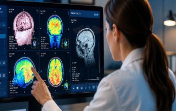
Follow-up combines neurological assessment with serial MRI. Treatment-related changes can increase contrast enhancement and sometimes resemble tumor progression. In a study of 169 patients with newly diagnosed glioblastoma who developed early radiographic progression after chemoradiation, 38.5% met the study’s clinical definition of pseudoprogression. A single early scan may therefore be insufficient, and clinicians consider symptoms, examination and changes on subsequent imaging [5].
Specialist Brain Radiation Therapy in Germany

Brain radiation in Germany is provided through specialist radiation oncology departments working with neuro-oncology, neurosurgery and neuroradiology. Multidisciplinary review is particularly important for complex cases and reirradiation. Modern external-beam techniques are available at specialist centres, while access to stereotactic and proton therapy varies between centres. The recommended technique and its availability should therefore be confirmed with the treating centre.
For international patients:
- Medical records, pathology and imaging can often be reviewed before travel.
- Previous radiation plans are reviewed where relevant.
- Aspecialist consultation assesses whether radiation may be appropriate.
- Preparatory planning may begin before arrival where possible.
For patients considering treatment from abroad, this often raises a practical question: How can I get a specialist opinion on brain radiation therapy in Germany?
A specialist review can assess the tumor type, location, previous treatment, imaging and treatment goals to help determine whether radiation therapy may be appropriate and which approach should be considered.
TIG GmbH (Treatment in Germany) can help international patients facilitate medical-record and imaging review, connect suitable cases with relevant radiation oncology specialists, and coordinate specialist consultations where applicable. The German medical team remains responsible for treatment assessment, recommendations and clinical care.
References
Why Patients Worldwide Prefer Our Medical Services in Germany – Key Benefits Explained
International patients planning breast cancer treatment in Germany may need support with hospital consultations, medical documentation, translations, and travel arrangements. TIG GmbH helps coordinate these practical steps and communication with healthcare providers according to each patient's needs and the services agreed.

The table above compares the support and coordination services offered through TIG GmbH with the arrangements patients may need to manage when booking directly with a clinic.





























.webp)
 (1).webp)

.webp)

.jpg)

 (1).webp)

.jpg)